Hyper IgE syndrome and eosinophilic ulcer
Vinitzky-Brener, Ilan1; Carrasco-Rueda, Carlos Alberto1; Ángeles-Gálvez, Mariana1; Alejandre-García, Alejandro1
Vinitzky-Brener, Ilan1; Carrasco-Rueda, Carlos Alberto1; Ángeles-Gálvez, Mariana1; Alejandre-García, Alejandro1
ABSTRACT
Hyper-IgE syndrome is a rare disorder characterized by elevated serum IgE, with skin manifestations (abscessed, eczema and infections) and recurrent pulmonary infections. This disorder occurs in two genetic patterns: Autosomal dominant (AD-HIES) and Autosomal recessive (AR-HIES). Eosinophilic ulcer of the oral mucosa is described as a benign, self-limited lesion with high predilection for the ventral mucosa of the tongue. Its diagnosis is established by histopathological study. We report the case of a 10-year-old male patient diagnosed with Hyper IgE Syndrome, with presence of Eosinophilic Ulcer in the oral mucosa, with approximately two months of evolution.KEYWORDS
Hyper IgE syndrome, eosinophilic ulcer, oral mucosa.Introduction
Hyper-IgE syndrome (HIES) is a multisystem disorder exhibiting immunologic and nonimmunologic features; the clinical triad that characterizes it is as follows: extreme elevations of serum IgE (higher than 2,000 IU/mL), cutaneous manifestations (eczema, abscesses or staphylococcal infections), and recurrent pulmonary infections. Staphylococcal skin abscesses may appear without signs of inflammation (cold) but are usually filled with pus; eczema may present as a newborn rash and is usually caused by Staphylococcus aureus; candidiasis may also manifest. Cases of pneumonia are complicated by parenchymal abnormalities of the lung (bronchiectasis and pneumatoceles).1,2
HIES was first described in 1966 by Davis SE et al.3 as "Job's syndrome", reporting that two girls showed severe eczema, pulmonary infections, and cold abscesses related to Staphylococcus aureus. In 1972, the syndrome was redefined by Buckley HR et al.,4 reporting two cases that also had similar infectious problems and a distinctive facial phenotype along with highly elevated serum IgE levels.
HIES is rare, with an annual incidence of 1:1'000,000 displaying no gender or racial differences. It's a genetic disease that can present two patterns:
The pathophysiology of the syndrome is still unknown; however, the etiology of the autosomal dominant form (AD-HIES) has been described as an error in the DNA binding domains and SH2 essential for the differentiation of Th17 cells, whose function involves the elimination of fungi and extracellular bacteria through the production of cytosines (IL-17, IL-22). The autosomal recessive form (AR-HIES) is probably due to a mutation present in the TYK2 gene, which emerges through B- and T-cell immunodeficiency.1,6
The diagnosis should be determined based on clinical and laboratory manifestations, mainly by identifying a genetic defect (STAT3 gene mutation); furthermore, the diagnosis can be established through the Grimbacher criteria (scoring system by the US Institutes of Health in patients with a family history of HIES). A score ≤ 30 has a sensitivity of 87.5% and specificity of 80.6% if less than 20 points are obtained: Dx unlikely; 20-40 points: Dx doubtful; and > 40 points: Dx probable. Differential diagnosis includes allergic bronchopulmonary aspergillosis, chronic granulomatous disease, T-cell lymphoma, and atopic dermatitis.7,8 In most patients with HIES, there are a series of oral manifestations; among them, there are anomalies in the process of exfoliation of the primary dentition in 64% approximately. This alteration results in the ectopic eruption of permanent teeth and the development of malocclusion. In more than 75% of people enduring this pathology, the oral mucosa and gingiva lesions have been identified, markedly affecting the dorsum of the tongue, palate, labial and buccal mucosae.2 In appearance, lesions on the palate are usually fibrous; abnormalities on the tongue appear in the form of grooves on the surface; lesions that develop at the mucosal level consist of shallow fissures and striae, patches, or plaques covered by a keratin layer, all of which are asymptomatic.2
Eosinophilic ulcer of the oral mucosa (EU), also known as traumatic ulcerative granuloma with stromal eosinophilia (TUGSE),9 is described as a rare, self-resolving, benign clinical-evolving ulcerative lesion, histologically composed of abundant eosinophils among the cells of the inflammatory infiltrate found in the dermis; its incidence is slightly more frequent in women.10,11
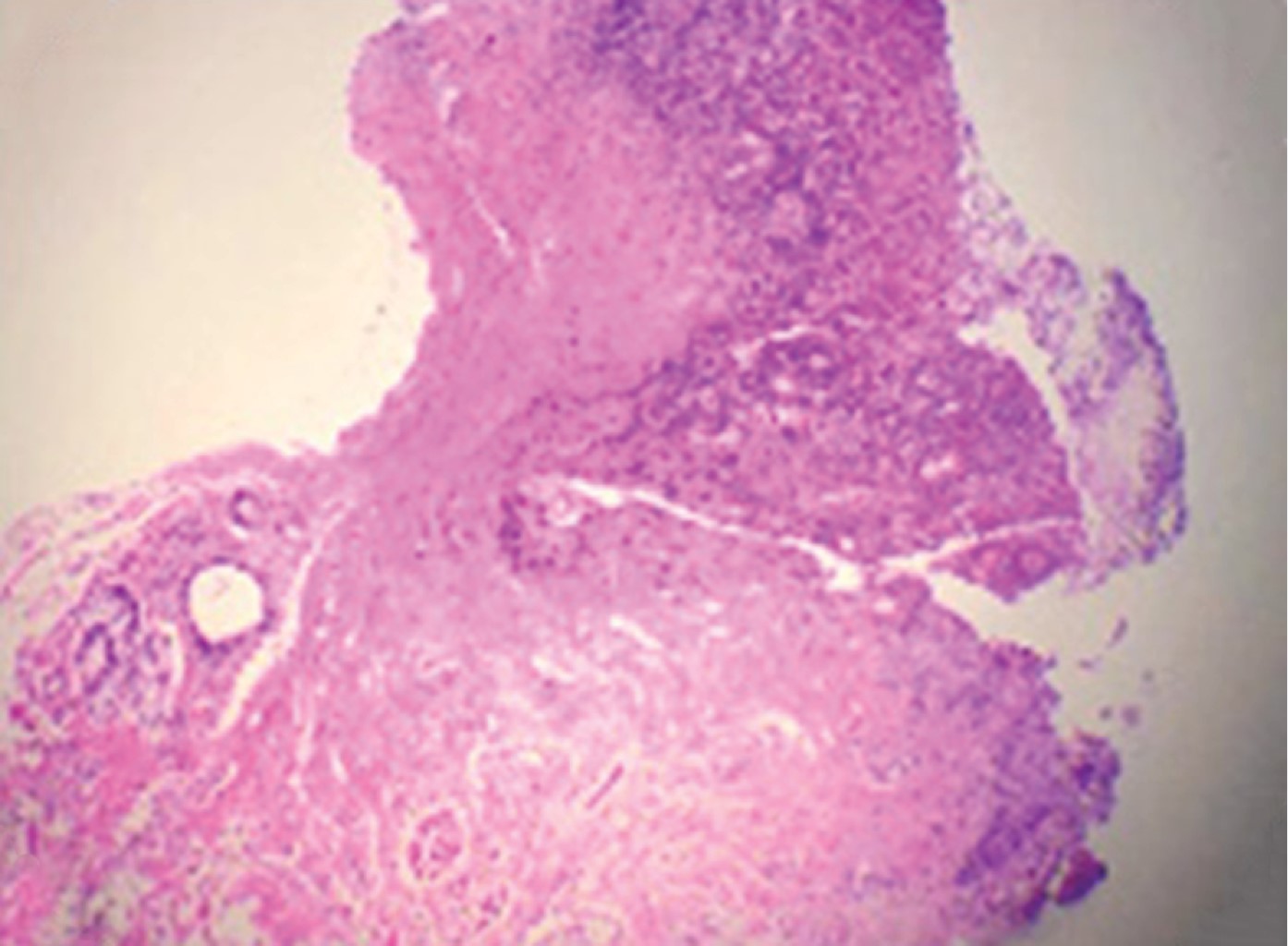
Clinically, it presents as an ulcer of 1 to 2 cm in diameter with indurated borders, asymptomatic, or extremely painful.12 Histologically, the ulcerated area of the mucosa appears covered by fibrinoid exudate with cellular detritus. At the base of the ulcer, there is granulation tissue and hyperplastic epithelial margins; the submucosa is occupied by a diffuse infiltrate composed of abundant eosinophils, lymphocytes, plasmacytes, and histiocytes.13,14
This article aims to present the case of a patient diagnosed with hyper-IgE syndrome and eosinophilic ulcer in the oral mucosa since we consider it relevant for pulmonologists to be aware of the possible oral manifestations of this condition, as well as for dentists to know about the hyper-IgE syndrome and its repercussions in the stomatognathic apparatus.
Clinical case
Here is the case of a ten-year-old male patient diagnosed with HIES, community-acquired pneumonia (CAP), and infectious exacerbation of bronchiectasis. A relative reported that the child had been admitted to the Pediatric Hospital two months before, seeking treatment for pneumonia and "a lesion in the mouth". Later, he was hospitalized at the Instituto Nacional de Enfermedades Respiratorias (INER), due to developing a productive cough having green, viscous, regular, non-cyanotizing, rubicundizing expectoration, frontal headache, preceded by nausea and dizziness for three days. The initial evaluation showed a Glasgow of 15, fever of 38.1 oC, RR: 38 rpm, HR: 154 bpm, BP: 110/60 mmHg, O2 saturation of 83% at room conditions, and serum IgE level of 4,570 IU/mL, general dehydration, referring pain at the abdominal level. A clinical-radiological diagnosis of pneumonia was made, and treatment with ceftriaxone and fluconazole was started empirically due to eosinophilia in peripheral blood.
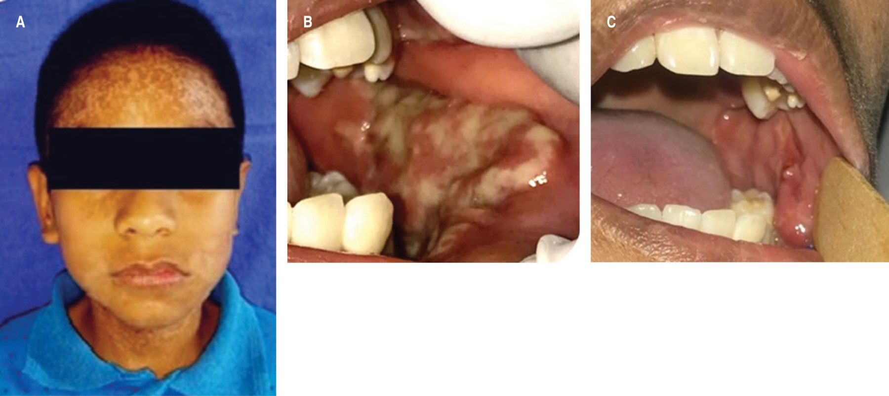
Facial examination shows lips and mucous membranes dehydrated, asymmetric auricular pavilions, hypochromic macules in the frontal and palpebral areas of the face, neck, and arms, and isochoric pupils. He also manifests the typical facial phenotype of the syndrome: broad nose, prominent forehead, facial asymmetry, and retention of some primary teeth (Figure 1A).
Intraoral examination reveals the presence of mixed dentition, multiple carious lesions, posterior open bite, and generalized gingivitis. In the left buccal mucosa, there was an ulcerated increase in volume extending from the labial mucosa to the cheek, of indurated consistency, covered by a greenish-yellow membrane and sessile base, for which the maxillofacial surgery department approached the patient (Figure 1B). An incisional biopsy was performed, evidencing an eosinophilic ulcer (Figure 2). The use of 0.20% chlorhexidine gel was indicated. Seven days after treatment, there was a notable improvement and decrease in the size and discomfort of the lesion (Figure 1C).
Discussion
Several authors have published texts on the oral implications associated with HIES, such as anomalies in the change of dentition, alterations in facial growth, and high susceptibility to oral infections; however, no cases reported in the literature were found in which EU is related to oral mucosa manifestations in patients having HIES. In this case, the patient demonstrated the main clinical and immunological characteristics of the syndrome. The physical features were coarse, prominent forehead, broad nose, dental retentions, a history of eczema, recurrent respiratory diseases (pneumonia and bronchiectasis), atopic dermatitis, oral herpes, generalized gingivitis, ulcers in the oral mucosa, and presence of vulgar flat warts. It was of fundamental importance to take a biopsy due to the appearance of the lesion to rule out the possibility of a malignant neoplasm.
Grimbacher B et al.14 suspected that the process of late rhizolysis of the primary dentition could be a manifestation of the same defect resulting in an ineffective inflammatory response. Bencini AC et al.13 describe that EU consists of a benign inflammatory process, characterized by a single ulceration with clear borders, and the causes that produce it in the oral mucosa are multiple: chemical, physical and thermal trauma; infectious agents (bacteria, viruses, fungi, parasites…); allergic reactions, systemic diseases; and lymphoproliferative disorders. In the case of our patient, we consider that the development of this type of non-neoplastic lesion is directly related to the high susceptibility to viral, bacterial, and fungal infections, among others; as a result of the error in the DNA binding domains and SH2 in the STAT3 gene, there is a defective inflammatory response against pathogens.
It was decided to perform conservative treatment with the application of 0.20% chlorhexidine gel, in addition to the placement and indication of the use of an occlusal guard; favorable results were obtained, without requiring any other therapeutic alternative, as indicated by Ficarra G et al.,15 who propose the use of antibiotics, cryosurgery, surgery, corticosteroids or surgery combined with intralesional corticosteroid therapy.
An accurate diagnosis of this pathology is of significant importance due to its multiple manifestations and clinical similarity with other entities that respond to different treatments and evolution. From the stomatological approach, the dentist's role in diagnosing and treating these lesions is highly relevant. The pulmonologist should be familiar with the oral implications that patients enduring HIES may present and refer them promptly to the dentist for evaluation and treatment.
AFILIACIONES
1Instituto Nacional de Enfermedades Respiratorias Ismael Cosío Villegas.Conflict of interests: The authors declare that they have no conflict of interests.
REFERENCES


