Clinical and chest imaging characteristic in patients with COVID-19 in a hospital from Tegucigalpa, Honduras
Cárcamo, Sandra1; Pacheco, Walter2,3; Ortiz, Glenda3,1; Rivera, María Félix1
Cárcamo, Sandra1; Pacheco, Walter2,3; Ortiz, Glenda3,1; Rivera, María Félix1
ABSTRACT
KEYWORDS
COVID-19, chest tomography, pulmonary patterns, CORADS RSNA.Introduction
The pandemic generated by the new coronavirus (COVID-19) since its initial emergence in late December 2019 in Wuhan City, China, has left 5.65 million deaths worldwide.1
The Hospital María de Especialidades Pediátricas is a national referral hospital that provides care for children under 18 years of age; however, due to the global emergency it joined the national hospital network for the care of patients with COVID-19. The World Health Organization (WHO) has a definition of a COVID-19 case, according to clinical manifestations and laboratory tests, suggested as follows: "confirmed case" person with reverse transcriptase polymerase chain reaction-reverse transcriptase (RT-PCR) test result positive for COVID-19, independent of signs and symptoms; "suspected case" patient with acute respiratory illness (fever and at least one symptom or sign of respiratory illness such as cough or dyspnea) and history of travel or residence in localities with reported community transmission during the 14 days prior to the onset of signs and symptoms. Also probable case: a suspected patient whose PCR-RT result is inconclusive or a suspected patient for whom laboratory testing could not be performed for any reason. In Honduras, the Ministry of Health approved the use of the antigen test for confirmation of COVID-19 according to the recommendations of the Pan American Health Organization (PAHO) and the Center for Communicable Diseases (CDC).2,3
This infection can present as mild, moderate or severe disease, including severe pneumonia, acute respiratory distress syndrome (ARDS), sepsis and septic shock, even leading to death.4
Computed tomography (CT) is considered the routine modality for the diagnosis, management and follow-up care of patients with COVID-19 pneumonia. It can aid in the early detection of pulmonary abnormalities for screening patients with suspected disease, especially patients with an initial negative RT-PCR screening result.5-7
By means of universal scales such as the Total Severity Score (TSS) and the Pulmonary Tomographic Assessment Tomographic Assessment Pulmonary Score System adapted to segmental involvement (PATPAS), a scale applied at the national level with which it is possible to know what percentage of the lung is affected by tomography.
The apropos of the study was to determine the clinical imaging features on chest CT in adult patients with COVID-19 at Hospital Maria de Especialidades Pediátricas, from April 2020 to April 2021.
Material and methods
Observational, cross-sectional study, with analytical component, conducted at the Hospital María de Especialidades Pediátricas from April 2020 to April 2021 located in the city of Tegucigalpa. There was a total population of 1,730 hospitalized patients, of which only 461 underwent institutional tomography, the sample was 149 patients who met the case definition.
Inclusion criteria were confirmed or probable COVID-19 patients older than 18 years who were hospitalized and underwent PCR-RT or antigen testing and high-resolution (HR) chest CT on the GE ligthspeed 64-slice facility CT scanner.
Procedure
The files of radiological studies and clinical records of patients admitted with a confirmed or probable diagnosis of COVID-19 who underwent tomography were reviewed, taking into account only those patients who presented tomographic findings typical of COVID-19, and then reevaluated together with the radiologist with four years of experience.
The findings were classified as follows: central (predominantly in the inner two thirds of the lung), peripheral (predominantly in the outer third of the lung) central and peripheral (in multiple lung segments) according to the European Society of Radiology and patchy distribution was added.8
To measure the degree of lung involvement by CT, the TSS was used, estimating the percentage of involvement in each lobe by the following score: 0 (none), 1 (affecting less than 5% of the lobe), 2 (affecting 5-25% of the lobe), 3 (affecting 26-49% of the lobe), 4 (affecting 50-75% of the lobe) or 5 (affecting more than 75% of the lobe). The CT score was obtained by summing the scores of the five lobes, for each patient the CT score was in the range of 0 to 25.9
In addition, two scales were applied to estimate the severity and suspicion of COVID-19: PATPAS of the Honduran Association of Radiology and Imaging (AHRI), and the Reporting and Data System for COVID-19 (CO-RADS) of the Radiological Society of North America (RSNA), respectively.10
PATPAS system: a five-point score was applied for each lung segment (0% no involvement, 25% mild, 25-50% moderate, 50-75% severe and more than 75% severe). CO-RADS provides a standardized system and evaluation scheme that simplifies reporting with a five-point scale of suspicion for lung involvement by COVID-19 on chest CT. CO-RADS 0: scans that are incomplete or of insufficient quality. CO-RADS 1: very low level of suspicion of lung involvement. CO-RADS 2: low level of suspicion of pulmonary involvement. CO-RADS 3: indeterminate findings of pulmonary involvement. CO-RADS 4: high level of suspicion of lung involvement by COVID-19 based on CT findings that are typical. CO-RADS 5: implies a very high level of suspicion of lung involvement based on typical CT findings. CO-RADS 6: was introduced to indicate COVID-19 proven by a positive RT-PCR test.11,12
Statistical analysis: univariate statistical analysis was performed (for qualitative variables; frequencies, proportions, 95% confidence intervals) and quantitative variables; means, mode, median, standard deviation. For bivariate analysis, Pearson correlation tests were used to see if the differences were statistically significant between the predominant pattern - evolutionary phase and the predominant pattern - disease severity variables.
The statistical programs used were SPSS, EPI INFO 7.2 and Excel for Windows.
Results
We evaluated 149 patients (77 women) with the most frequent age range between 40-59 years of COVID-19 patients, with a mean of 56 years. The most frequently found antecedents were hypertension (69 [46.3%]), obesity (49 [32.9%]) and diabetes/prediabetes (23 [15.4%]). Clinical manifestations at admission were dyspnea (132 [93.3%]), fever/febrile (124 [83.2%]) and cough (109 [73.2%]) (Table 1).
Patients were hospitalized between a most frequent range of one to seven days with a mean interval of 1.5 (0.643 standard deviation), of which 28 (18.8%) were admitted to ICU (intensive care unit), deceased patients were 17 [11.4%].
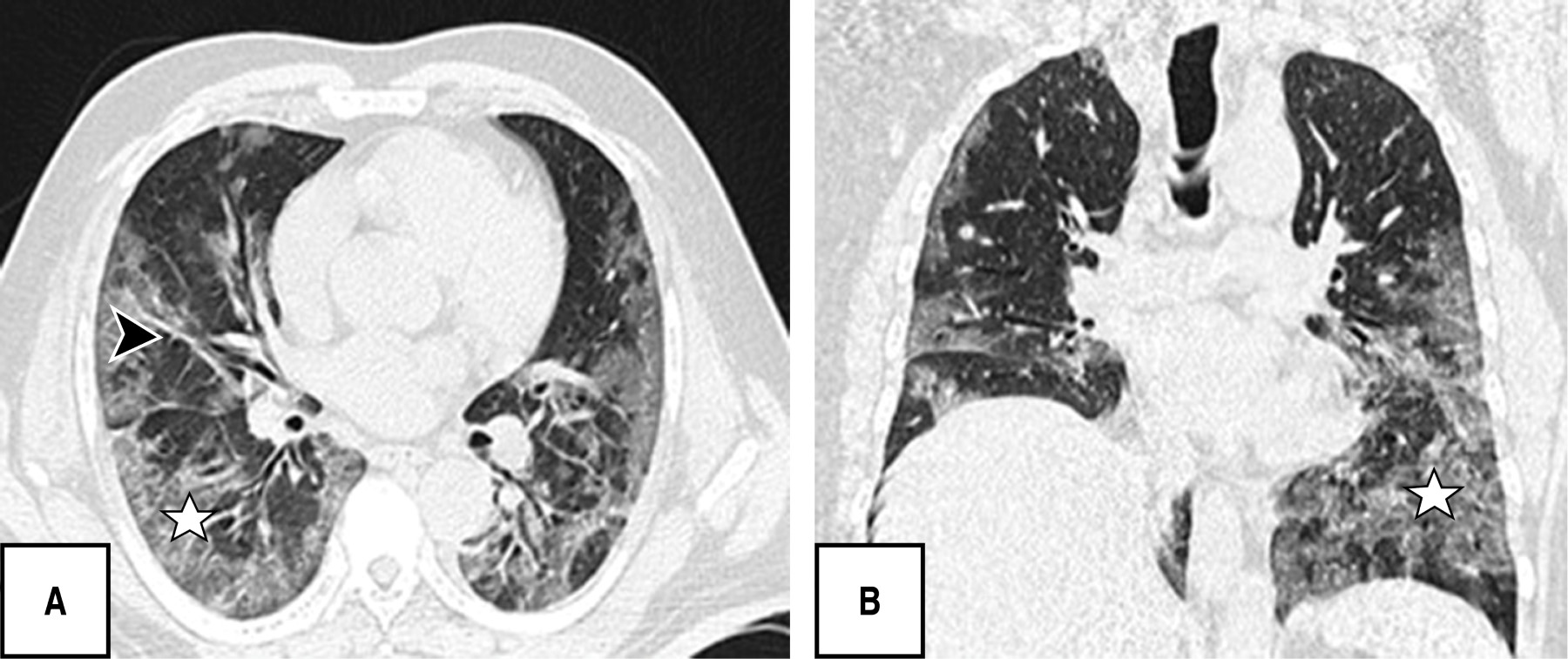
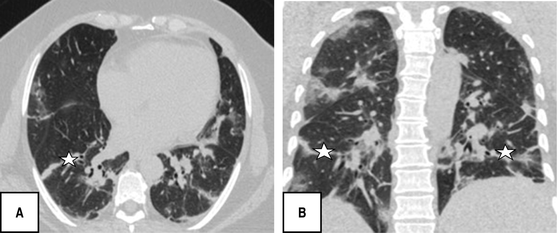
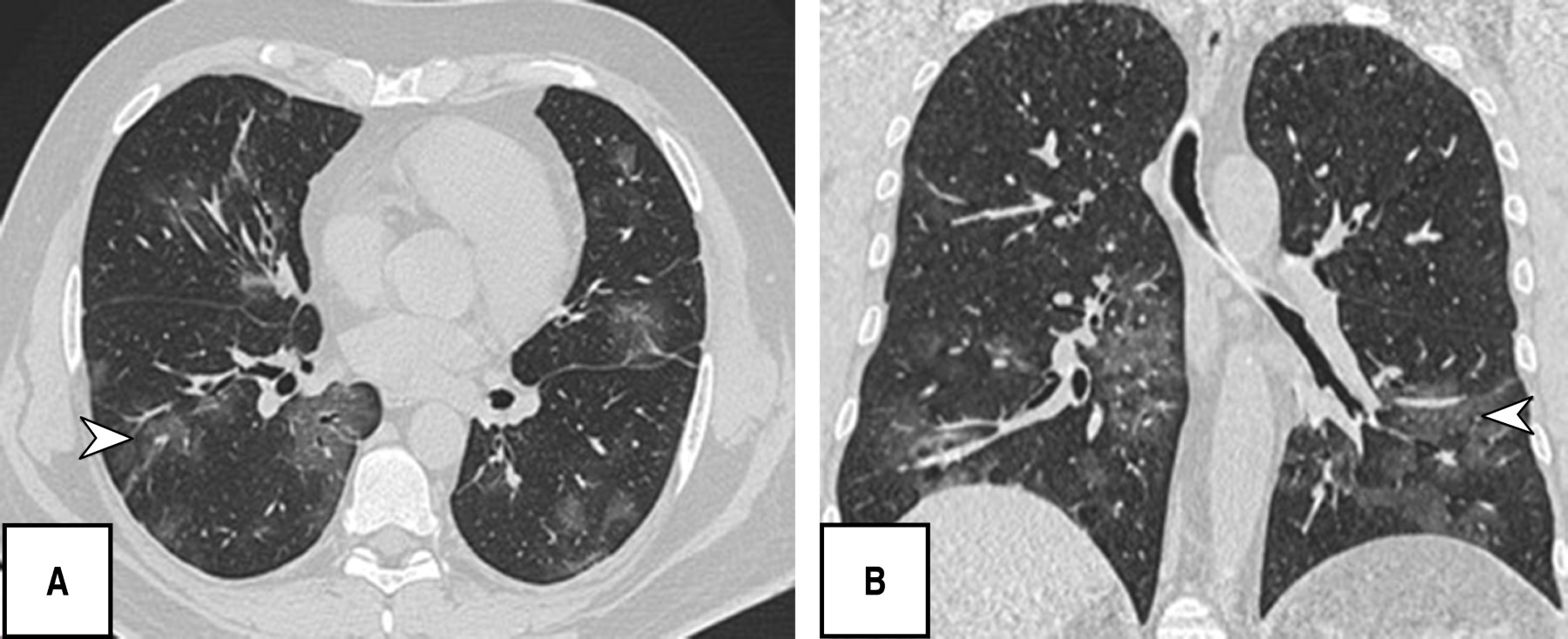
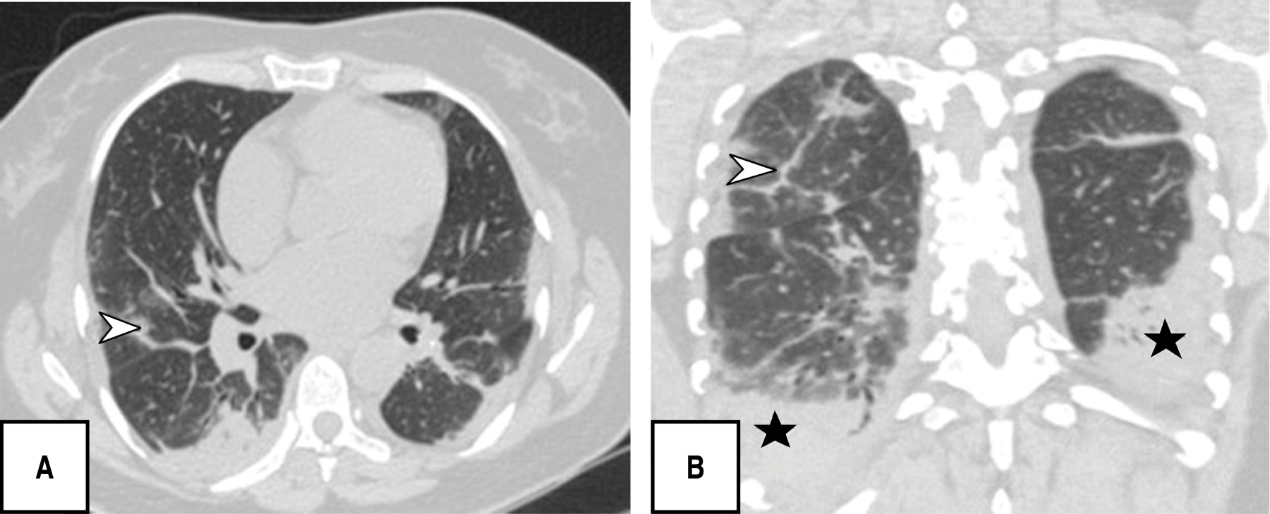
During the tomographic evaluation, the most frequent pulmonary patterns were: cobblestone pattern (120 [80.5%]), pleuroparenchymal bands (118 [79.2%]) and ground glass pattern (110 [73.8%]), reticular pattern (82 [55%]), and in lower percentage vascular dilatation (56 [37.6%]) and consolidated pattern (52 [34.9%]) (Figures 1, 2, 3 and 4).
Regarding the phases found, the resorption phase was observed most frequently (69 [46.3%]) followed by the peak phase (40 [26.8%]) (Table 2).
Each patient was found to have five of five lung lobes affected, 144 (96.6%), with a total percentage of involvement of 26-49%, (96 [62.4%]); the most characteristic distribution of lung involvement was central and peripheral (86 [57.7%]), and without segment predominance (86/57.7%) followed by localization in posterior segments (59/39.6%), all with statistical significance (p < 0.05). The PATPAS severity score presented by the patients was moderate (93 of 149 patients [62.4%]).
Eighty-six patients were identified with positive laboratory tests by PCR-RT, which were included within the CORADS 6 category (cases already confirmed by laboratory plus typical finding by CT), in addition patients confirmed by antigen test were included, thus adding up to a total of 143 out of 149 (93%); only six patients had a negative laboratory test by PCR-RT and antigen, which were categorized as CO-RADS 5 with very high suspicion of presenting the disease by typical findings by CT AR (p < 0.05).
The Pearson correlation (0.65) obtained with a null probability (0%) of being random allows us to conclude that there is a high positive correlation between the predominant pulmonary patterns and the phase of tomographic evolution.
Discussion
A study conducted in Colombia by Marrin Sanchez stated that this disease occurs more frequently in the male sex at a mean age of 65.75 ± 18.1. In this study the female sex was more frequent and the age ranged between 40-59 years of age with a mean of 56 years. However, a study carried out by PAHO states that the incidence between 40-59 years of age is equal in both sexes, and as age increases, it is more frequent in men.13,14
The most frequently found personal pathologic antecedents were hypertension, obesity and diabetes/prediabetes (similar to the study by Peña et al. where hypertension ranked first), as well as that presented by Murrieta et al. where patients showed to have two or more comorbidities.15,16
In our study, the initial clinical manifestations that prevailed were dyspnea (132 [93.3%]), fever/febrile fever (124 [83.2%]) and cough (109 [73.2%]), a finding similar to that of Song et al. observing fever (49 of 51, 96%) and cough (24 of 51, 47%) among the most frequent symptoms, as well as a retrospective study in patients hospitalized in the ICU, where they reported that the most common initial symptoms were fever (92%), cough (68%) and dyspnea (49%).17,18
It is noteworthy that most of the patients in this research presented in a chronic/advanced phase of the disease, finding as predominant patterns the cobblestone (120/149; 80.5%), pleuroparenchymal bands (118/149; 79.5%) and in third place ground-glass pattern (110/149; 73. 8%), without identifying any case with findings of atoll, pneumothorax or twinning tree, other studies do report ground glass opacities as the predominant finding followed by the consolidation pattern and in this regard pleural effusion, pericardial effusion, lymphadenopathy, cavitation, the reverse halo or atoll sign and pneumothorax are rare, but can be observed with the progression of the disease.19-21
Segmental involvement was without predominance and with a central and peripheral distribution in equal percentage with 57.7%, different to that published by Pan F et al., where the lower lobes were more likely to be involved and in general, the subpleural distribution of lesions was more frequent than central lung lesions.22
Soriano et al. reported that the presence of ground-glass opacities, reticular pattern, cobblestone pattern, subpleural lines, pleural thickening and fibrosis were more frequently found in the intermediate/progressive phase, especially in the advanced phase; similarly we observed a significant correlation (Pearson's index of 0.65) between the predominant pattern and the progressive phase of the disease.23 The CT scores of the progressive stage group were significantly higher than those of the early stage group; however, in this study there was no statistical significance between CT pattern and severity score.
Conclusions
Recognizing the evolutionary phases of COVID-19 disease will help us to provide adequate follow-up and to know the patient's prognosis, for which the chest tomography complemented with the PCR-RT laboratory test is the diagnostic basis when undertaking the management.
The Hospital María de Especialidades Pediátricas as a national reference hospital during the pandemic did a great job, assisting adults with COVID-19 in the face of a national problem, thus obtaining complete and valuable information for the present study.
The study was limited in some deceased patients because the laboratory test was not presented in the file, since it was necessary even if it was negative or positive.
AFILIACIONES
1Universidad Nacional Autónoma de Honduras 2Hospital María de Especialidades Pediátricas 3Instituto Hondureño de Seguridad Social.Conflict of interests: the authors of this study declare that they have no conflict of interests.
REFERENCES
Secretaría de Salud, Honduras) _Lineamientos para el uso de la prueba rápida de detección de antígenos para la COVID-19 resolución No. 33 DGN- DEC19-21:2020 del 6 de noviembre del 2020. [Internet] (Citado el 10 de agosto 2021) Disponible en: http://www.salud.gob.hn/site/index.php/component/edocman/sesal lineamientos-para-el-uso-de-las-pruebas-ra-pidas-de-deteccio-n-de-antigeno-para-covid-19-2
Rubin G, Ryerson C, Haramat L, Sverzellati N, Kanne J, Raoof S, et al. The role of chest imaging in patient management during the COVID-19 pandemic: a multinational consensus statement from the fleischner society. Chest Journal. Radiology. 2020;296:172-180. Available in: https://doi.org/10.1148/radiol.2020201365
Simpson S, Kay F, Abbara S, Bhalla S, Chung J, Chung M, et al. Radiological Society of North America Expert Consensus Document on Reporting Chest CT Findings Related to COVID-19: Endorsed by the Society of Thoracic Radiology, the American College of Radiology, and RSNA. Radiology: Cardiothoracic Imaging. 2020;2(2):200152. Available in: https://doi.org/10.1148/ryct.2020200152
Equipo del Sistema de Gestión de Incidentes (IMST) / Oficina de Equidad, Género y Diversidad Cultural (EGC) diferencias por razones de sexo en relación con la pandemia de covid-19 en la región de las américas de enero del 2020 a enero del 2021_OPS [Internet] [Citado 25 junio 2021] Disponible en: https://www.paho.org ' file '
Soriano Aguadero I, Ezponda Casajús A, Mendoza Ferradas F, Igual Rouilleault A, Paternain Nuin A, Pueyo Villoslada J, Bastarrika G, et al. Hallazgos en la tomografía computarizada de tórax en las fases evolutivas de la infección por SARS-CoV-2. Radiología. 2021;63(3):218-227. Disponible en: https://doi.org/10.1016/j.rx.2021.02.004


|
Table 1: Epidemiological-clinical characteristics in patients with COVID-19. N = 149. |
||
|
General data |
n (%) |
p (χ2) |
|
Gender Female Male |
77 (51.7) 72 (48.3) |
0.000
|
|
Median age 56 years 20-39 40-59 60-79 80 years and more |
25 (16.8) 64 (43.0) 47 (31.5) 13 (8.7) |
0.000*
|
|
Procedence Tegucigalpa, Francisco Morazán |
149 (100.0) |
|
|
Pathological antecedents Hypertension Obesity Diabetes mellitus/prediabetes Heart disease Asthma Alcoholism and smoking Neoplasia COPD Nephropathy Other antecedents Hypothyroidism |
69 (46.3) 49 (32.9) 23 (15.4) 11 (7.4) 10 (6.7) 7 (4.7) 2 (1.3) 2 (1.3) 1 (.7)
11 (7.4) |
0.066 0.514
0.145 0.559 0.034 0.388 0.388 0.624
0.004 |
|
Clinical manifestations Dyspnea Febrile fever Cough Myalgia Headache Arthralgia Anosmia Ageusia Odynophagia |
139 (93.3) 124 (83.2) 109 (73.2) 36 (24.2) 33 (22.1) 31 (20.8) 25 (16.8) 22 (14.8) 22 (14.8) |
0.441 0.306 0.427 0.059 0.196 0.118 0.476 0.276 0.063 |
|
Other symptoms Diarrhea Rhinorrhea |
17 (11.4) 19 (12.8) |
0.000 0.000 |
|
* Values obtained for Student’s t. Source: data obtained from the archive of the Hospital María de Especialidades Pediátricas |
||
|
Table 2: Lung patterns by AR tomography in patients with COVID-19. N = 149. |
|||
|
Tomographic patterns |
n (%) |
χ2 |
p |
|
Tarnished glass pattern Consolidated pattern Cobblestone pattern Reticular pattern Pleuroparenchymal bands Bronchial thickening Vascular dilatation Pleural effusion Adenopathies Atelectasis Others (granuloma) |
110 (73.8) 52 (34.9) 120 (80.5) 82 (55.0) 118 (79.2) 10 (6.7) 56 (37.6) 10 (6.7) 10 (6.7) 44 (29.5) 22 (14.7) |
0.017 1.581 0.221 1.170 0.958 0.263 4.460 1.413 3.477 0.294 15.549 |
0.521 0.140 0.395 0.181 0.219 0.441 0.025 0.200 0.057 0.359 0.000 |
|
CT disease stage Initial Progressive Peak Resorption |
11 (7.4) 29 (19.4) 40 (26.9) 69 (46.3) |
7.152 21.682 32.925 77.384 |
0.004 0.000 0.000 0.000 |
|
CT = computed tomography. HR = high resolution. Source: data obtained from the archive of the Hospital María de Especialidades Pediátricas. |
|||


