Chest X-ray in pediatrics. A systematic interpretation
Aguilar-Viveros, Brenda1; Zamora-Ramos, Margarita2; Martínez-Martínez, Blanca Estela1; Thomé-Ortiz, Laura Patricia1
Aguilar-Viveros, Brenda1; Zamora-Ramos, Margarita2; Martínez-Martínez, Blanca Estela1; Thomé-Ortiz, Laura Patricia1
ABSTRACT
Reading the chest radiograph requires training and repetition. The basic projections are: left lateral and posteroanterior. It is recommended to follow an order in interpretation: patient identification, projection, description of the technique (using the mnemonics GERICA), review of soft tissues, bony parts, diaphragmatic dome, mediastinum, cardiac silhouette and finally the lung parenchyma; all this considering the differences in pediatric age (changes in the technique by age group, bone variations, normal values of the cardiothoracic index, presence of thymus, etc.). All this will help us not to overlook any alteration and get the most out of this study, which is present in the daily life of every clinician.KEYWORDS
chest radiograph, pediatrics, interpretation.Introduction
Chest radiography is a usual diagnostic instrument in pediatric patient care that provides timely information, hence the importance of proper analysis and interpretation. It is commonly said, "A picture is worth more than a thousand words". An adequate exercise in radiographic evaluation can provide dynamic information derived from a static image.1
Radiological technique
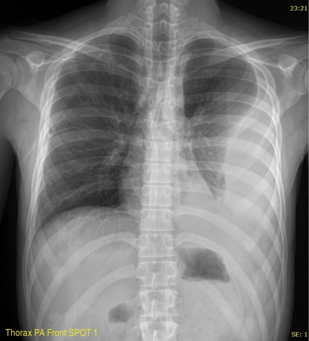
The routine radiological examination of the thorax makes two projections: posteroanterior (PA) and left lateral (L). They are performed with the patient standing or sitting, the PA at a focus-film distance of 1.8 meters, and the left side at 2 meters.
The correct exposure is achieved using kilovoltage peaks (kVp), ranging from 100 to 140, to reduce the radiation scatter index, visualize the bone structures with better density and, in turn, adequately identify the lung parenchyma and mediastinum. The image results from the sum of the passage of a polychromatic X-ray beam through an object that contains areas with different absorption coefficients, registering on the film the combination of the response to the intensity of the emitted light and the non-absorbed portion of radiation.1-4 They are performed in maximum inspiration and in apnea.
In the chest X-ray, the mediastinal structures and the diaphragm overlap the lung parenchyma, so in a PA projection, we will have 40% hidden areas; therefore, a lateral projection is always recommended.2-4
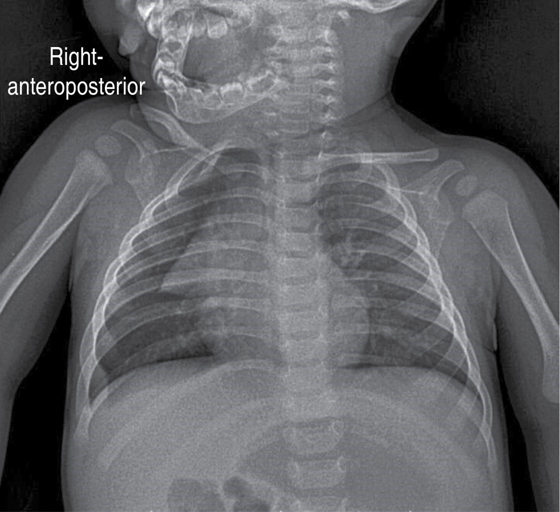
In patients with poor clinical conditions, infants, and uncooperative patients, frequent in the pediatric age, X-rays are taken in an anteroposterior projection (AP) and with portable equipment, with the drawback of obtaining images of low technical quality; since this projection is performed at a shorter focus-film distance, structures are magnified, and fewer sharp images are obtained.5
There are other radiological projections, such as expiratory radiography, which detects small pneumothoraxes and localized air trapping associated with foreign bodies, and the lateral decubitus projection with a horizontal beam that evaluates free fluid in the pleural cavity, among others.
Basic concepts and terminology
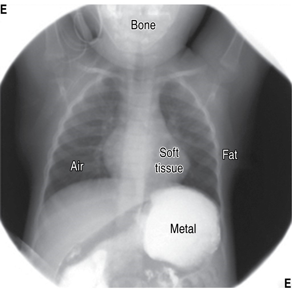
In conventional radiology, we have five densities: air, fat, water, calcium, and metal (Figure 1).
Remember, radiolucent is any object, organ, or tissue that allows light to pass through it and translates as dark shades. While radiopaque is any object, organ, or tissue that does not allow the passage of light or puts resistance to the passage of light on it and translates as light shades in the radiological image.
Interpretation
There should be a systematized analysis in the interpretation of thoracic radiography. We suggest the following order:
All radiological projections, including the lateral one, are interpreted in the same order.
We propose the GERICA mnemonic to remember them during the interpretation.
Radiological description
It is suggested to start with the parts from the least to the most interesting to avoid overlooking minor alterations.
Direct
Indirect
Conclusions
Chest radiography is a particularly useful tool in the study of many pediatric pathologies and is part of the initial evaluation. Its proper interpretation can be the starting point in the suspected diagnosis. It is essential that it is performed systematically and that the differences inherent to the pediatric age are considered.
AFILIACIONES
1Centro Médico Nacional Siglo XXI, Instituto Mexicano del Seguro Social, Mexico CityConflict of interests: the authors declare that they have no conflict of interests.
REFERENCES
Guzzo-De León D. Análisis secuencial segmentario para el diagnóstico de cardiopatías congénitas. El aporte de la radiología, del electrocardiograma y de la ecocardiografía. Rev Urug Cardiol [Internet]. 2008;23(1):21-48. Disponible en: http://www.scielo.edu.uy/scielo.php?script=sci_arttext&pid=S1688-04202008000100004&lng=es


|
Table 1: Cardiothoracic index (CT) values at pediatric ages.9-11 |
||
|
Age |
CT index |
Range |
|
0-3 weeks |
0.55 |
0.45-0.65 |
|
4-7 weeks |
0.58 |
0.46-0.70 |
|
1 year |
0.53 |
0.45-0.61 |
|
1 a 2 years |
0.49 |
0.39-0.60 |
|
2-6 years |
0.45 |
0.40-0.52 |
|
7 years |
< 0.50 |
0.40-0.50 |


