Tracheal lipoma: an unusual case
Midence-Arguello, María José1; Morales-Gómez, José1; Solares-Espinoza, Andrea Gloria1; Vázquez-Minero, Juan Carlos1
Midence-Arguello, María José1; Morales-Gómez, José1; Solares-Espinoza, Andrea Gloria1; Vázquez-Minero, Juan Carlos1
ABSTRACT
Tracheal lipoma is a rare, benign tumor with a very low incidence, whose symptoms are non-specific and variable according to the degree of airway obstruction, causing unnecessary morbidity and mortality if not adequately treated. We describe a case of tracheal lipoma with obstructive airway symptoms in a 70-year-old patient, with symptoms of six months' evolution, productive cough and progressive dyspnea which limited his daily activity. He presented with stridor, without data of respiratory distress, bilateral rales; complementary studies were carried out to rule out tracheal stenosis due to clinical symptoms. The tomography showed an adherent encapsulated lesion in the anterolateral tracheal wall with significant airway obstruction of 95%. The treatment of choice was endoscopic resection using laser or cryotherapy. Interventional treatment via bronchoscopy should only be applied to narrow-based benign tracheal tumors that protrude into the tracheal lumen. In our case, the decision was made to resect using this technique. Follow-up at one month without recurrence, adequate airway patency, and resolution of symptoms prior to the procedure.KEYWORDS
lipoma, tumor, endotracheal, bronchoscopy, case report.Introduction
Tracheal primary tumors are rare. In adults, most of them are malignant; only 10-20% of tracheal tumors are benign, including chondromas, papillomas, fibromas, hemangiomas, lipomas.1-4 Tracheal lipoma is an extremely rare benign neoplasm whose incidence ranges from 0.1 to 0.5%; the symptomatology and signs are nonspecific, such as dry cough, wheezing and sometimes shortness of breath. Due to the slow-growing characteristic, patients with benign tracheal tumors develop symptoms of airway obstruction gradually. They are almost always misdiagnosed as asthma, chronic obstructive pulmonary disease or bronchitis. Definitive diagnosis of these tumors is often delayed.1-3 They require resection and can be approached endoscopically as well as by open surgery. We present a patient with obstructive symptoms due to the presence of an endotracheal tumor, treated with rigid bronchoscopy and resection.
Presentation of the case
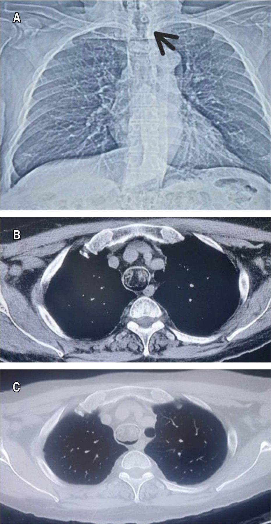
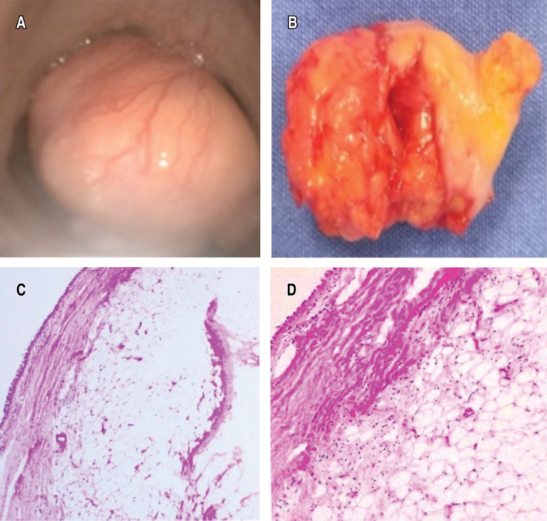
A 70-year-old man, with clinical picture of six months of evolution, characterized by dyspnea of medium efforts, progressing to small efforts and productive cough, with hyaline expectoration, not cyanosing or dyspneic. During the physical examination, vital signs: blood pressure (BP) 113/101 mmHg, HR 79 bpm, RR 22 rpm, temperature 36.2 °C, oxygen saturation 91% room air, initial arterial blood gasses pH 7.45, PCO2 36.7 mmHg, PO2 65 mmHg, with stridor, no evidence of respiratory distress, bilateral rales were auscultated in the lung fields. Oxygen was administered through nasal prongs at two liters with oxygen saturation at 94%. Chest X-ray was performed without parenchymal alterations, with an image of tracheal stenosis (Figure 1A). A simple chest CT scan was requested, showing an irregular encapsulated lesion adhered to the anterolateral wall of the middle third of the trachea (Figures 1Band 1C). Fibrobronchoscopy was performed (Figure 2A), and a 3 × 2 cm whitish tumor was identified at the level of the sixth tracheal ring obstructing 95% of its pedunculated tracheal lumen in the anterior wall. Resection was performed by rigid bronchoscopy and total resection of the tumor (Figure 2B). Histopathological examination reported tracheal lipoma (Figures 2C and 2D). The patient presented good evolution with no data of respiratory compromise. He was discharged due to improvement, with tomographic follow-up one month after the procedure without evidence of recurrence, with adequate patency of the tracheal lumen.
Discussion
Most primary tracheal tumors in adults are malignant and benign tumors are rare. Primary benign tumors of the trachea include mainly leiomyoma, papilloma, fibroma, hemangioma, chondroma, and mixed salivary gland tumors; whereas lipoma is very rare.1-4
Airway lipomas affect the main bronchi and less frequently the trachea, as in our case. They usually originate in the submucosal fat of the tracheobronchial tree, are usually pedunculated, may extend between cartilage rings into peritracheal tissues, and recur after resection.1
Benign tracheal tumors tend to grow slowly and are asymptomatic in the early stages. They are difficult to diagnose during this period; moreover, they are misdiagnosed after respiratory symptoms such as bronchial asthma or pulmonary infections appear. Symptoms of airway obstruction appear when the degree of tracheal blockage increases from 50 to 75% or when the luminal diameter is less than 8 mm. In our case, the patient presented dyspnea.1-3,5 He had a clinical history of six months of evolution with respiratory symptoms and dyspnea on exertion. The clinical picture is compatible with lesions or tumors occupying the tracheal lumen as in this case.
In patients with gradually worsening dyspnea it is suggested to consider the possibility of a tracheal tumor, it is necessary to perform a timely examination; including thoracic computed tomography (CT) and bronchoscopy can provide an accurate diagnosis. In fact, the sign of this disease is mainly inspiratory dyspnea, and it is evidently different from expiratory dyspnea caused by bronchial asthma and pulmonary infectious diseases. A careful physical examination may be helpful in the differential diagnosis.
Commonly, it is difficult to find a lesion on a conventional chest radiography because the trachea is covered by the mediastinum, almost always seen as normal, as in this case (Figure 1).1-3 Studies such as CT and flexible bronchoscopy are valuable for diagnosis, which were performed in our case.1-3
CT of the neck and chest showed a soft tissue tumor endotracheally and in the anterior wall of the tracheal wall. Resection was adequately performed without complications, especially since pathologic biopsy reported a benign tracheal lipoma. Endoscopic treatment by loop resection or cryotherapy is recommended as the first approach.
Interventional treatment through bronchoscopy should only be applied to benign tracheal tumors with a narrow base, originating from submucosal, nodular or polypoid tissues protruding into the tracheal lumen, where the base is connected to the tracheal wall by a pedicle of irregular length, where the tissue surface and irrigation is smaller and, therefore, the risk of bleeding decreases. In our case, the decision was made to resect it by this technique.
Tracheal resection with reconstruction should be considered when the histopathological study provides evidence of malignant margins or the radiological image shows tumor extension through the tracheal wall and endoscopic treatment fails.1-3,5
Conclusions
Most tumors of the tracheobronchial tree are malignant. Benign tumors are rare and lipoma is extremely rare as in our case. Tracheal lipoma is histologically benign and may cause airway obstruction. Primary tracheal tumors should be suspected in patients with recurrent dyspnea symptoms and respiratory symptomatology. Cervical and thoracic CT scans should be considered; flexible bronchoscopy should be timely, as it can provide an accurate diagnosis.
Interventional treatments through fibrobronchoscopy are mainly suitable for benign tracheal tumors with narrow margins. However, if bronchoscopic treatment is not possible, surgical resection should be considered to decrease the mortality risk of this pathology.1-3,5
AFILIACIONES
1Instituto Nacional de Enfermedades Respiratorias Ismael Cosío Villegas. Mexico City, Mexico.Conflict of interests: the authors declare that they have no conflict of interests.
REFERENCES


