Sex-associated differences in pulmonary disease caused by mycobacteria diagnosed at INER from 2016-2018
Sartillo-Mendoza, Luis G1,3; Martínez-Sanabria, Claudia A1,3; Becerril-Vargas, Eduardo2; González, Yolanda2; Juárez, Esmeralda2
Sartillo-Mendoza, Luis G1,3; Martínez-Sanabria, Claudia A1,3; Becerril-Vargas, Eduardo2; González, Yolanda2; Juárez, Esmeralda2
ABSTRACT
KEYWORDS
mycobacteria, pulmonary disease, tuberculosis, non-tuberculous mycobacteria.Introduction
Pulmonary tuberculosis, mainly caused by Mycobacterium tuberculosis (M. tuberculosis), is the second leading cause of death associated with a single pathogen in the world. In 2021, men accounted for 56%, women 32% and children12% of new global cases; in addition, 53% of deaths recorded during 2021 occurred in men, 31% in women and16% in children.1 Men, on the other hand, are more susceptible than women to developing severe forms with greater lung damage, have greater need for retreatment, more co-infections and, in some countries with high incidence, are more susceptible to developing drug resistance.2-4
Non-tuberculous mycobacterial lung disease (NTM) presents with a clinical picture similar to that of tuberculosis and can be caused by a variety of mycobacterial species.5 This disease is geographically diverse and the existing data are heterogeneous due to the lack of national coverage reports, but, in general, it is accepted that the majority of cases occur by species members of the M. avium complex (M. avium and M. intracellulare).6 In the United States and Canada, the majority of cases occur in women (86% and 61%, respectively), although population-based studies are not reported, nor is an association of sex and age with mortality reported.7,8
The predominance of either sex in tuberculosis and mycobacterial lung disease could have an effect on clinical practice, follow-up of cases to prevent complications, and personalized supervision of drug therapy. In addition, the difference between the sexes makes it necessary to implement new clinical and basic research protocols for the identification of socioeconomic and immunological determinants of susceptibility. Therefore, this work aims to identify whether there is a predominance of one sex in lung disease caused by M. tuberculosis and other mycobacteria in a third-level Mexican hospital.
Material and methods
Description of the study and origin of the data. This study is a retrospective case series. It consisted of searching for cases with lung disease that have tested positive for any mycobacteria during the years 2016 to 2018 in the archives of the Clinical Microbiology Service of the National Institute of Respiratory Diseases Ismael Cosío Villegas (INER) of Mexico City. The study was approved by the INER science and research ethics committees (code C56-22) with a waiver of the letter of informed consent by virtue of the fact that the research was retrospective, the confidentiality of the information was ensured and the personal identification of the patients was eliminated.
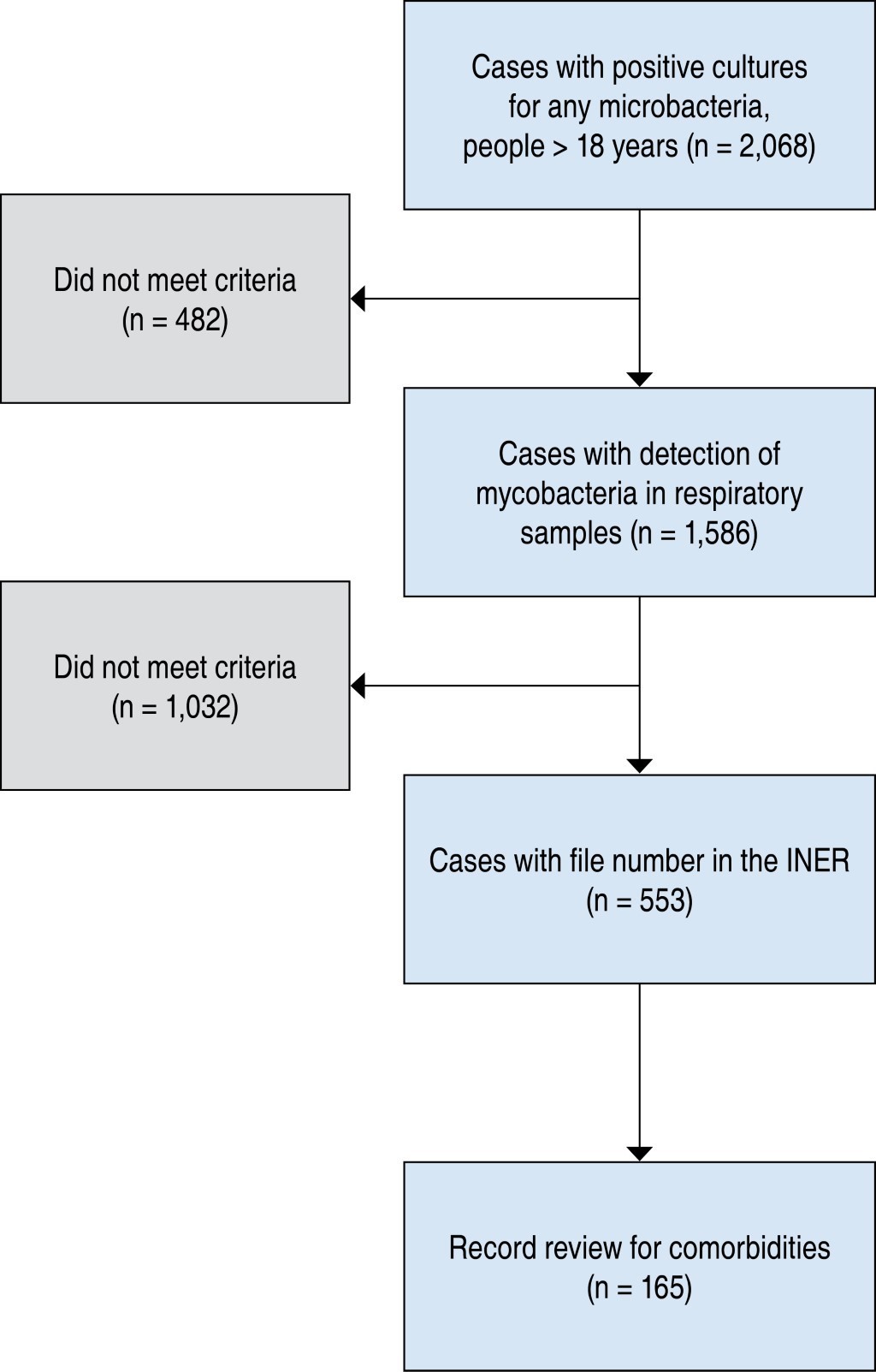
Selection of the study sample. In the file of the Clinical Microbiology Service, 2,068 case records with positive culture for any mycobacteria were found. All patients whose samples were of pulmonary origin such as expectoration, bronchoalveolar lavage, lung biopsy and bronchial aspirate from individuals over 18 years of age who had a record in the INER were selected for this study. A total of 553 patients were included (Figure 1). For the analysis of comorbidities, a random selection of 165 files was made, 81 men and 84 women. The number of positive cases for the different complexes was taken into account in the calculation of the sample size of each group.
Statistic analysis. The number of individuals with lung disease caused by the different mycobacteria is presented as number of individuals or percentage. These data are shown in tables or proportion graphs and are only described, no statistical tests were performed. To establish the differences in continuous variables in the different types of mycobacteria, the Kruskal-Wallis test was performed, followed by the Dunn test for comparisons of pairs of samples. Categorical variables are presented in tables. Men and women were compared for each type of mycobacterium using Fisher's exact test. In all cases, p < 0.05 was considered significant and the GraphPad Prism ver 9 software (La Joya, CA, USA) was used.
Results
Mycobacteria found in samples of respiratory origin in adults. This study was conducted retrospectively covering the years 2016, 2017 and 2018. The samples used for diagnosis were expectoration (50.63%), bronchoalveolar lavage (31.1%) and lung biopsy (18.26%). M. tuberculosis is the mycobacterium that occurred in the largest number of cases in samples of respiratory origin (Table 1). Mycobacteria M. avium, M. bovis and M. intracellulare were the mycobacteria other than M. tuberculosis that were found in the highest number of cases. Other mycobacteria occurred less frequently.
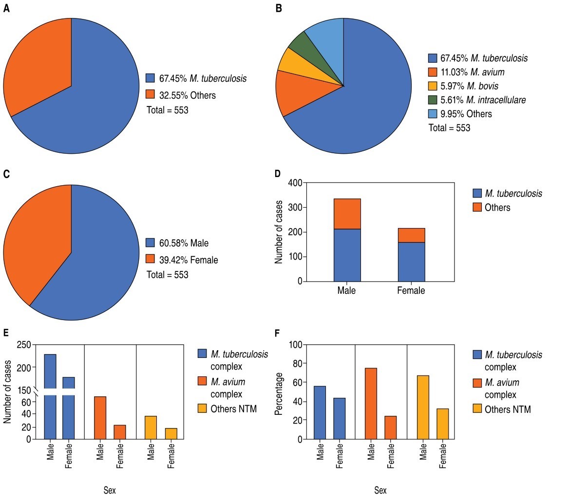
Distribution by sex of mycobacterium species reported in the study period. Pulmonary tuberculosis was the main pulmonary disease caused by a mycobacterium (67% of cases); the next in frequency were M. avium, M. bovis and M. intracellulare (among the three represent 22.61%), and the other mycobacteria represent 9.95% (Figure 2A and 2B ). 60.58% of patients were male (Figure 2C) and these comprise the largest number of cases with M. tuberculosis and with any other mycobacterium (Figure 2D). Because M. tuberculosis and M. bovis belong to the M. tuberculosis complex (M. tuberculosis complex: M. tuberculosis, M. bovis, M. africanum, M. canetti, M.microtti) and M. avium and M. intracellulare belong to the M. avium complex, the data were grouped into three categories, the M. tuberculosis complex, M. avium complex and other mycobacteria. The highest number of cases are men in the three groups, M. tuberculosis complex (56.40%), M. avium complex (75%) and other NTM (62.79%) (Figure 2E and 2F ).
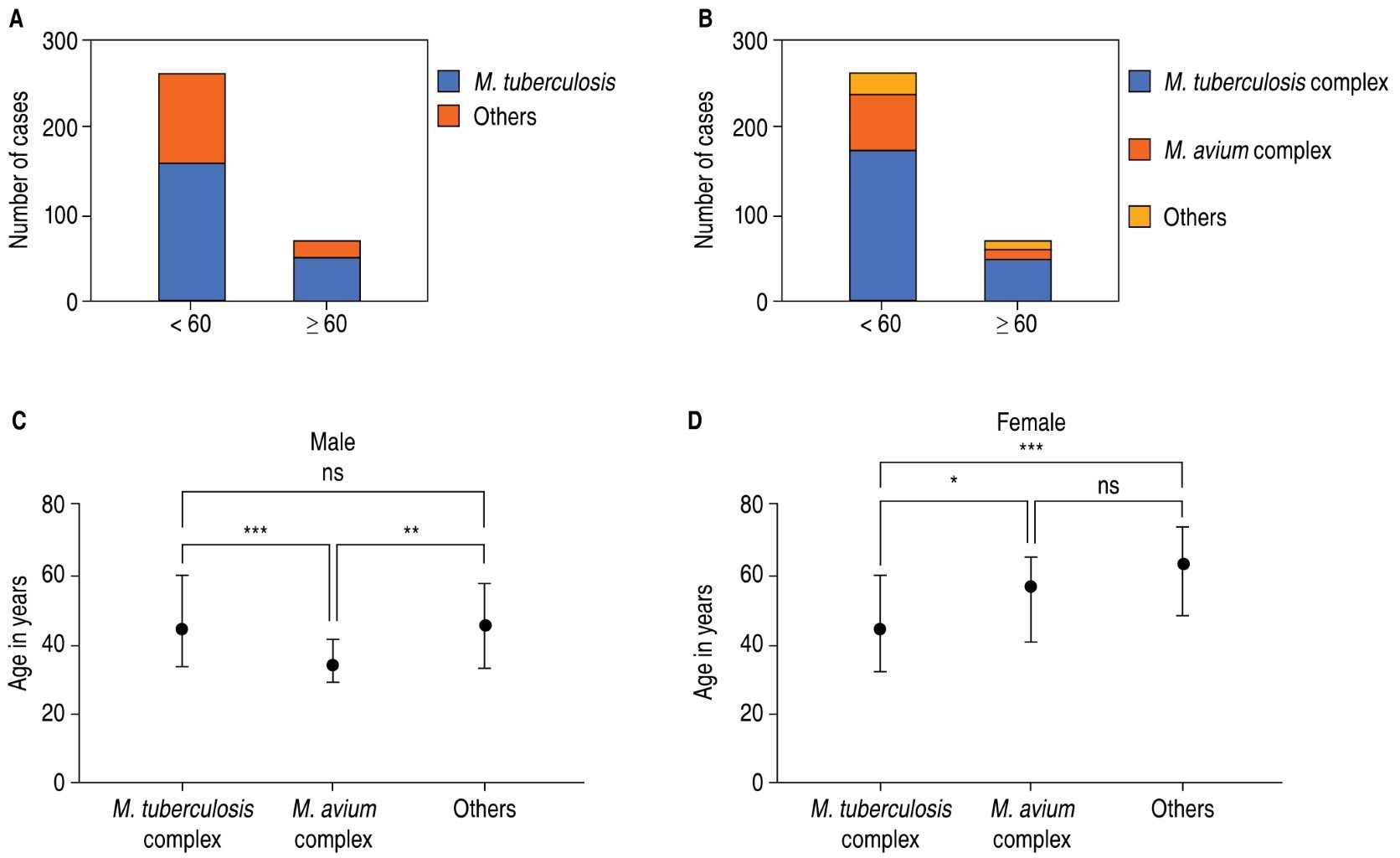
Additionally, we observed that the majority of patients were younger than 60 years of age (Figure 3A), and the pattern is preserved when separated by mycobacterial complex (Figure 3B). There was no difference in the ages of men and women infected by members of the M. tuberculosis complex. In the male group, patients infected by members of the M. avium complex were significantly younger than those infected by M. tuberculosis and other MNT, but there was no difference between the latter two (Figure 3C). Women who became infected with members of the M. avium complex and other NTM were significantly older than those who became infected with members of the M. tuberculosis complex (Figure 3D).
Main comorbidities observed in men and women with lung disease caused by mycobacteria. To determine the relevance of comorbidities, 81 men and 84 women were randomly selected and analyzed by group (Table 2). The proportion of patients with human immunodeficiency virus (HIV) was significantly higher in men infected with members of the M. tuberculosis and M. avium complexes, but not in the other NTM; this directly influenced the overall. The proportion of women with hypertension was significantly higher in patients infected with members of the M. avium complex. There were no differences between men and women, in either group, in the proportion of individuals with diabetes mellitus or cancer.
Discussion
Mycobacterial infections are a growing global health problem. Both M. tuberculosis and NTM infection are often seen in people who have a compromised immune system, which not only makes infection permissible, but also favors the development of active lung disease.9,10 In this study we investigated the prevalence of the different species of mycobacteria in patients who come with lung disease with suspected tuberculosis to a Third Level Respiratory Disease Hospital, the INER, who had a positive culture for any species of mycobacteria, and identified whether there is a predominance of one sex in lung disease caused by mycobacteria.
We observed that the lung disease under study was mainly caused by M. tuberculosis. This infection occurred more frequently in men under 60 years of age and was associated with a greater number of comorbidities, the main one being diabetes mellitus. These data are consistent with other studies, in which a higher prevalence has been reported in men.11,12 The most frequent NTMs included mycobacteria of the M. avium and M. bovis complex, which are part of the M. tuberculosis complex. For this reason, we divided the groups into those infected by members of the two complexes and by other NTM.
Mycobacterial infections of the M. avium complex, which includes M. intracellulare, are the most common NTM found in our study group. These data are also consistent with what was recorded in other populations.10,13,14 Contrary to expectations, we also observed this infection predominantly in men. Unlike infections by members of the M. tuberculosis complex, we detect that infection caused by members of the M. avium complex is more frequent in people under 35 years of age, perhaps associated with HIV infection. In other populations it has been recognized that M. avium infection occurs mainly in people over 65 years of age.13 This phenomenon is only observed in women. We also observed that the prevalence of M. avium in people living with HIV is higher than that reported in other populations,15 so M. avium monitoring is required in this specific group of patients.
Unlike reports of M. tuberculosis infections, which occur similarly in most populations, the prevalence of infections by members of the M. avium complex depends on the populations studied.16 We observed that infection by members of the M. avium complex in the group of women occurs in older than 60 years, with hypertension and diabetes mellitus as the main comorbidities. There are no reports of association between M. avium infection and hypertension, although M. avium has been reported concomitantly with acute kidney disease as a rare clinical event.17 Additionally , susceptibility to M. avium infection has been reported in postmenopausal women with low levels of dehydroepiandrosterone sulfate (DHEA-S), without alterations in estrogen levels (estradiol and estrone);18 but other reports suggest that this susceptibility is associated with low levels of estradiol (E2), without alterations in DHEA-S,19 so more studies are required to elucidate the role of hormones in susceptibility to M. avium infection in women.
We also observed that infections with other NTM such as M. abscessus, M. chelonae, M. fortuitum, M. gordonae and M. simiae occurred more frequently in men under 60 years of age. Contrary to what has been reported in other countries, these infections were less frequent in women and occurred mainly in those over 50 years of age. Although not significantly, these NTM occurred in people with exposure to wood smoke, smoking, or drug use.
Some immune functions are associated with sex. For example, steroid hormones influence the production, maturation, differentiation, and function of immune system cells and molecules.20 Estradiol and progesterone decrease the expression of proinflammatory cytokines, such as IL-1β, IL-6, IL-2,TNF-α , and IFN-γ , and influence the production of lipid mediators of inflammation resolution.21-23 On the other hand, the X chromosome has genes associated with the regulation of the immune response, such as those encoding IL-2, TLR7, TLR8, IL-1, IRAK1, the IL-3 and IL-13 receptor alpha chain, and several small RNAs (miRNAs) regulating the immune response, while the Y chromosome has none.21,24,25 These features likely promote less severe clinical pictures in women. Future research will determine the contribution of these factors to mycobacteriosis.
This study has limitations mainly related to bias in the selection of samples. First, the INER is a National Institute of Health, a Third Level Hospital that mainly treats complicated or severe lung diseases. Second, because this is a retrospective study, the universe of samples that were evaluated were only those cases in which there was a clinical record in the INER. However, the result is relevant to propose additional analyses, epidemiological studies and new protocols for personalized care.
Conclusions
In a Third Level Care Center, men are mainly affected with lung infections caused by mycobacteria, both M. tuberculosis and the other NTM. There are currently no treatment policies that consider the predominance of men in pulmonary mycobacteriosis. Due to its impact on personalized medicine, the search for biomarkers and the follow-up of treatment, it is necessary to evaluate the phenomenon at the national level, and find out what are the social and biological factors that determine it.
AFILIACIONES
1Facultad de Medicina, Benemérita Universidad Autónoma de PueblaConflict of interests: the authors declare that they have no conflict of interests.
REFERENCES
Chidambaram V, Tun NL, Majella MG, Ruelas Castillo J, Ayeh SK, Kumar A, et al. Male sex is associated with worse microbiological and clinical outcomes following tuberculosis treatment: A retrospective cohort study, a systematic review of the literature, and meta-analysis. Clin Infect Dis. 2021;73(9):1580-588. Available in: https://doi.org/10.1093/cid/ciab527
Danley J, Kwait R, Peterson DD, Sendecki J, Vaughn B, Nakisbendi K, et al. Normal estrogen, but low dehydroepiandrosterone levels, in women with pulmonary Mycobacterium avium complex. A preliminary study. Ann Am Thorac Soc. 2014;11(6):908-914. Available in: https://doi.org/10.1513/AnnalsATS.201312-422OC
Marriott I, Bost KL, Huet-Hudson YM. Sexual dimorphism in expression of receptors for bacterial lipopolysaccharides in murine macrophages: a possible mechanism for gender-based differences in endotoxic shock susceptibility. J Reprod Immunol. 2006;71(1):12-27. Available in: https://doi.org/10.1016/j.jri.2006.01.004


|
Table 1: Species of mycobacteria found in samples of respiratory origin between 2016 and 2018 in the INER. |
||||
|
Species |
Number of cases |
|||
|
2016 |
2017 |
2018 |
Total |
|
|
M. tuberculosis |
99 |
127 |
147 |
373 |
|
M. avium |
17 |
22 |
22 |
61 |
|
M. bovis |
7 |
9 |
17 |
33 |
|
M. intracellulare |
12 |
11 |
8 |
31 |
|
M. abscessus |
2 |
6 |
7 |
15 |
|
M. chelonae |
3 |
2 |
3 |
8 |
|
M. fortuitum |
1 |
1 |
4 |
6 |
|
M. gordonae |
1 |
3 |
2 |
6 |
|
M. simiae |
1 |
3 |
2 |
6 |
|
M. kansasii |
0 |
3 |
1 |
4 |
|
M. genavense |
0 |
1 |
1 |
2 |
|
M. mucogenicum |
0 |
1 |
1 |
2 |
|
M. szulgai / M. lentiflavum |
0 |
0 |
1 |
1 |
|
M. malmoense |
0 |
0 |
1 |
1 |
|
M. bovis / M. chelonae |
0 |
0 |
1 |
1 |
|
M. tuberculosis / M. chelonae |
0 |
1 |
0 |
1 |
|
M. scrofulaceum |
1 |
0 |
0 |
1 |
|
M. xenopi |
1 |
0 |
0 |
1 |
|
Total |
|
|
|
553 |
|
Table 2: Proportion of patients* with comorbidities by group. |
||||||||
|
Characteristics |
M. tuberculosis complex |
M. avium complex |
Others |
Total |
||||
|
Sex |
Male n = 55 |
Female n = 66 |
Male n = 19 |
Female n = 10 |
Male n =7 |
Female n = 8 |
Male n = 81 |
Female n = 84 |
|
Age, years, median (range) |
36.5 (20-85) |
34.5 (18-88) |
34 (21-54) |
56.4 (22-77) |
55 (28-65) |
60 (39-74) |
45 (20-85) |
45 (18-88) |
|
HIV‡ |
12 (21.8)§ |
3 (4.5) |
15 (78.94)¶ |
2 (20) |
4 (36.3) |
0 (0) |
31 (38.3)¶ |
5 (6) |
|
Cancer‡ |
0 (0) |
2 (3.03) |
1 (5.26) |
0 (0) |
0 (0) |
0 (0) |
1 (0.12) |
2 (2.4) |
|
Diabetes mellitus‡ |
21 (38.1) |
25 (37.8) |
0 (0) |
3 (30) |
0 (0) |
2 (25) |
21 (26) |
30 (35.7) |
|
Hypertension‡ |
8 (14.5) |
10 (15.1) |
0 (0) |
5 (50)§ |
0 (0) |
2 (25) |
8 (9.9) |
17 (20.2) |
|
Others (exposure to wood smoke, smoking, drug use) |
21 (38.2)§ |
9 (13.6) |
5 (26.3)§ |
0 (0) |
6 (85.7) |
5 (62.5) |
32 (39.5)|| |
14 (16.7) |
|
* Random selection of a sample of 81 men and 84 women. ‡ «Yes», frequency and percentage [n (%)]. § p > 0.05. ¶ p < 0.001. Fisher’s exact test || p < 0.01. |
||||||||


