Mediastinal cryobiopsy: case report
Bautista-Méndez, Rafael1; Montero-Reyes, Fernando1; Magdaleno-Maldonado, Gerardo Ezequiel1; Pineda-Gudiño, Rey David1
Bautista-Méndez, Rafael1; Montero-Reyes, Fernando1; Magdaleno-Maldonado, Gerardo Ezequiel1; Pineda-Gudiño, Rey David1
ABSTRACT
Bronchoscopy has become an essential diagnostic and therapeutic modality for a variety of lung diseases. With the addition of complementary techniques, such as taking a biopsy with a cryogenic probe, the role of the evaluation of pulmonary and mediastinal pathology is further expanded, allowing better diagnostic performance.KEYWORDS
mediastinal cryobiopsy, flexible bronchoscopy, transcarinal.Introduction
Flexible bronchoscopy has become an essential diagnostic and therapeutic modality for a variety of lung diseases; the addition of transbronchial needle aspiration (TBNA) further expanded the role in the evaluation of mediastinal pathology. In 1949, Schieppati made the first description of mediastinal lymph node sampling through the carina using a rigid bronchoscope. In 1978, Wang and colleagues demonstrated that paratracheal lymph node sampling by TBNA was feasible.1
Bronchoscopic cryobiopsy has proven useful in both endobronchial and peripheral lung tumours as well as interstitial lung diseases; the most common side effects reported are pneumothorax and bleeding. Mediastinal cryobiopsy has shown improved diagnostic utility for molecular testing of genetic mutations.2
The first randomized trial, conducted in 2021 by Zhang and associates, included a total of 197 cases with mediastinal lesions ≥ 1 cm in which they used endobronchial ultrasound (EBUS)-guided TBNA and linear EBUS-guided cryobiopsy, alternating the order of initiation of these procedures. They recorded higher diagnostic yield in cryobiopsies 91.8% versus 79.9% (p = 0.001) and higher sensitivity in rare tumours (91.7% versus 25%, p = 0.001), with pneumothorax and pneumomediastinum in 1% and 0.5% of cases, respectively, resolving without intervention.3-5
In another prospective pilot trial, conducted in 2022 by Gershman E et al, 24 patients underwent EBUS-guided cryobiopsy following EBUS-guided TBNA. They obtained an anatomopathological result of 83.3% for cryobiopsy and 87.5% for TBNA. No complications were recorded in any of the patients.4
In both studies mentioned above, the procedures were performed under EBUS guidance. However, in the present study, the procedure was performed without ultrasound guidance because the lesion was of significant size and located in a relatively easily accessible site.
The performance of the first mediastinal cryobiopsy at our hospital is described below.
Presentation of the case
Male patient aged 63 years, with no history of cancer in direct family members, smoking rate of 50 packs/year, suspended three months prior to hospital admission, recently diagnosed with systemic arterial hypertension.
His condition began three months prior to admission with dysphagia, for which he stopped smoking without improvement; he subsequently presented with non-productive cough and dysphonia. He went to the corresponding health centre where extension studies were performed, which revealed a mediastinal mass. He was referred to the third level for further treatment.
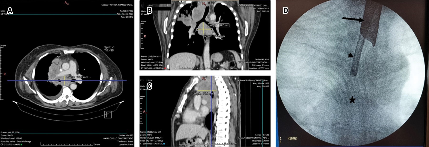
On admission, a simple and contrasted chest CT scan was performed; it showed a subcarinal mediastinal lesion measuring 58 × 51 × 74 mm, as well as a mass in the apical segment of the right upper lobe, a centrilobular micronodular pattern in the right upper and middle lobe, a subsolid nodule measuring 4. 3 × 5.2 mm in S7 and soft nodule of 5.4 × 5.1 mm in S8 of the right lower lobe, so it was decided to perform bronchoscopy with biopsy (Figure 1A-D). The procedure was performed under full sedation with the use of rigid tracheoscopy, with a rigid Hemer Richar Wolf® tracheoscope model with a diameter of 14 mm, and flexible bronchoscopy with a 5.9 mm diameter vi deobronchoscope with a 2.8 mm working channel Olympus Medical Systems®. With fluoroscopy support, a 21G eXcelonTM Boston Scientific® transbronchial aspiration needle was introduced for aspiration at the level of the main carina. Subsequently, Radial JawTM 4 Boston Scientific® forceps, 100 cm in length, with 1.8 mm diameter forceps were introduced with a double purpose: first, to collect tissue sample and, second, to increase the diameter of the orifice; finally, through the same puncture site, a flexible cryoprobe of 1. 9 mm × 900 mm Erbe® flexible cryoprobe connected to the ERBECRYO 2® cryosurgery unit, with a freezing time of four seconds, was thawed in warm saline; a total of three samples were collected, with no post-procedure bleeding. Two tissue samples, each measuring 0.1 × 0.1 cm, were obtained using forceps and three samples were obtained by cryobiopsy, together measuring 0.6 × 0.3 × 0.2 cm, ideal for immunohistochemistry and mutational studies in pathology.
The samples were sent to the Pathology Service for review by a pulmonary pathologist for immunohistochemistry and mutational studies. The report indicated the following:
Discussion
Cancer treatment is evolving rapidly; therefore, bronchoscopists are providing increasing amounts of tissue to perform molecular testing for genetic mutations in a safe and minimally invasive manner. The implementation of these tests has led to improved diagnostic and therapeutic performance without the need to subject patients to surgical procedures.
Surgical mediastinoscopy is still considered a first-line procedure in certain situations, such as in cases of haematological malignancy, and in conditions where there is failure of bronchoscopic specimen collection. However, the complication rate is higher and the scarce availability of ultrasound bronchoscopic guidance at the institutional level in our country makes this procedure a minimally invasive diagnostic option. In the case presented, we dispensed with the use of EBUS due to the size (> 5 cm) and location (subcarinal level) of the lesion, with adequate results. It is necessary to continue performing this technique in order to standardize it and to know the profile of patients who would benefit from this procedure.
Conclusions
Rapid and correct diagnosis of mediastinal masses is mandatory for the clinical management and prognosis of patients; to this end, it is necessary that sufficient high-quality tissue samples are obtained for pathological, genetic, immunological and other evaluations, with increasingly less invasive methods. Mediastinal cryobiopsy is a promising technique for obtaining biopsies with sufficient material for pathological analysis.
AFILIACIONES
1Hospital Central Militar. Mexico City, Mexico.Conflict of interests: the authors declare that they have no conflict of interests.
REFERENCES


