Sternoclavicular joint osteomyelitis as a cause of descending necrotizing mediastinitis
Hernández-Ramírez, Israel1; Espinoza-Mercado, Fernando1; Corona-Perezgrovas, Miguel Ángel1; Rivera-Navarrete, Eric1
Hernández-Ramírez, Israel1; Espinoza-Mercado, Fernando1; Corona-Perezgrovas, Miguel Ángel1; Rivera-Navarrete, Eric1
ABSTRACT
Descending necrotizing mediastinitis is a rare entity with a high morbimortality; the most common non-surgical causes are infections coming from oral or cervical region that spread through cervical fascias to mediastinum. However, there are other uncommon etiologies that affects by proximity mediastinum as sternoclavicular joint infection does; sternoclavicular joint infections (SCJI) constitute less than 1% of all joint infections and even 0.5% in healthy patients. We describe the case of a female 76 years old, diabetic patient with descending necrotizing mediastinitis associated with left SCJI, who unfortunately died, despite treatment provided. Likewise, a review of the current literature is made due to low frequency of this entity as a cause of mediastinitis.KEYWORDS
descending necrotizing mediastinitis, osteomyelitis, septic arthritis, sternoclavicular joint, case report.Introduction
Descending necrotizing mediastinitis is an urgent condition associated with high morbidity and mortality, and therefore requires a multidisciplinary approach. There are several classifications for the diagnosis and management of mediastinitis, one of the most popular is the one by Endo,1 who proposed that according to the tomographic extension it should be classified as type I when it is located in the superior mediastinum above the tracheal bifurcation, which may not require aggressive mediastinal drainage; type IIA when it extends to the lower anterior mediastinum and type IIB when it also includes the posterior mediastinum, requiring complete mediastinal drainage. The most common cause is an infection from the oral or cervical region that spreads through the fasciae to the superior mediastinum.
Treatment depends largely on the etiology, as well as the extent of the disease; in Mexico, an extensive review of this condition was carried out, seeking to standardize treatment from the surgical point of view, for a condition that represents a health problem in certain segments of our population.2 However, there are less common causes that affect the mediastinum by proximity, such as an infection in the sternoclavicular joint; this type of infection constitutes less than 1% of all joint infections. Due to the uncertain nature of its presentation, as well as its low prevalence, the diagnosis of a sternoclavicular joint infection is often delayed.3 Suppurative infections involving this joint are especially difficult to treat because of its proximity to major vascular structures and the absence of a sufficient amount of peripheral tissue to limit damage. Patient-specific factors as well as infection by resistant microorganisms often complicate the picture;4 because of these factors and the low incidence of such infections, surgical treatment has not been standardized.
We present the case of a patient with descending necrotizing mediastinitis associated with infection of the left sternoclavicular joint, as well as a review of the reported literature.
Case presentation
We present the case of a 76-year-old woman with long-standing diabetes with irregular insulin-based control, systemic arterial hypertension treated with captopril and losartan, as well as chronic renal failure treated with peritoneal dialysis; she was admitted to the emergency department by her relatives, with an approximate evolution of three days with general malaise, progressive neurological deterioration and abdominal pain. Based on the patient's medical records, she was approached with suspected dialysis catheter-associated peritonitis, meeting clinical and biochemical criteria of systemic inflammatory response, with a partially compensated metabolic acidosis with lactate of 6.1 mmol/L.
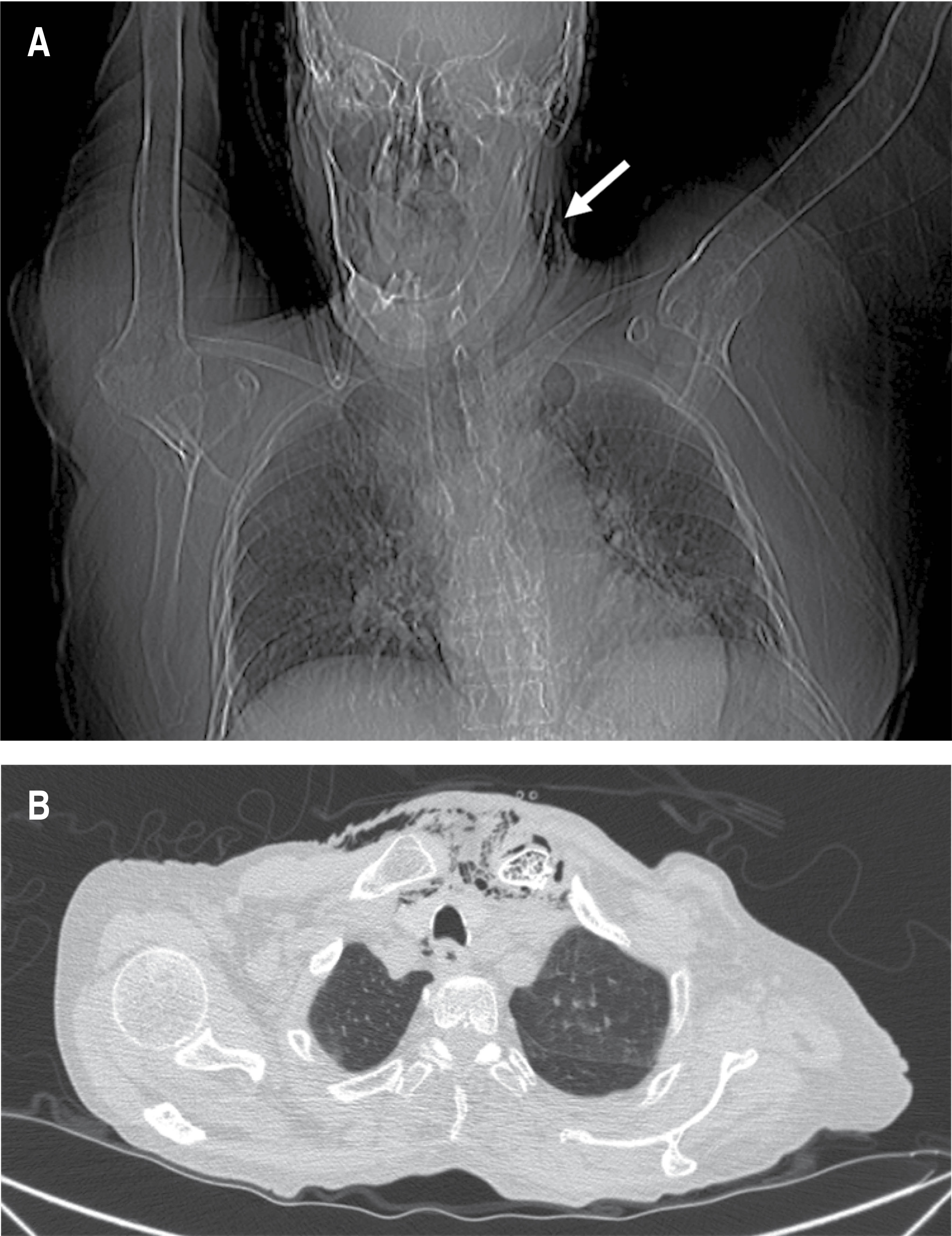
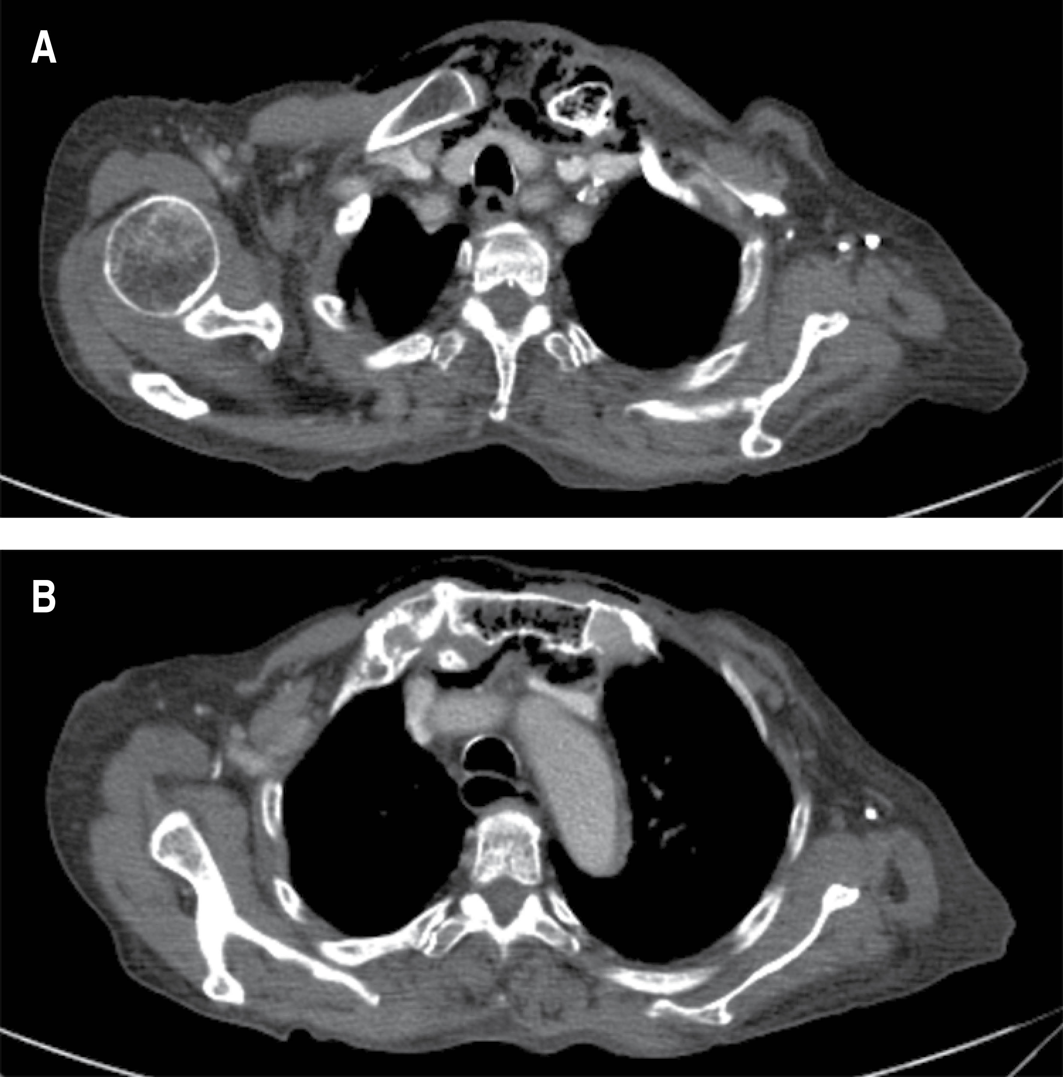
During the initial approach in the emergency department, it was decided to place a urinary catheter (the patient still had spontaneous diuresis), with evidence of pyuria; an attempt was made to place a central venous access, palpating the presence of subcutaneous emphysema, so it was requested to perform a computed tomography scan; with report of an inflammatory process located in the cervical region with extension to the upper mediastinum, as well as the presence of intramedullary gas in the left clavicle and sternal manubrium, suggestive of osteomyelitis (Figures 1 and 2). The study of the peritoneal fluid obtained through the dialysis catheter was found to be within reference parameters, so in view of the evidence of septic shock associated with radiological evidence of Endo IIA mediastinitis, urgent surgical treatment consisting of exploration and cervical drainage was proposed, which was not accepted by the patient's relatives until 12 hours after hospital admission. Broad-spectrum antibiotic treatment (carbapenemics) was started from hospital admission and during surgery a cervical collar approach was performed, 1 cm above the sternal notch, dissected in planes and the presence of purulent fluid from the left sternoclavicular joint was identified; Due to the hemodynamic instability of the patient, manifested by persistent hypotension, use of pressor amines with progressive doses and presence of ventricular extrasystoles, it was decided to limit surgery to temporary control of the infectious focus with drainage, irrigation and partial debridement of the affected area.
The patient was admitted to the Intensive Care Unit post-surgery, with a torpid evolution and progressive deterioration despite the established medical-surgical treatment; she died eight hours after surgery. The final report of the culture and Gram stain of peritoneal fluid requested was negative for the presence of microorganisms, urine culture with growth of more than three Gram (-) microorganisms, bone culture and cervical abscess positive for K. pneumoniae.
Discussion
Descending necrotizing mediastinitis is an urgent condition that demands an opportune diagnosis with aggressive treatment. Although there are frequent and well-established causes of this entity, infection of the sternoclavicular joint is not one of them, with few reports in the literature. Infection of this joint is rare, but when it does occur, it results in abscess formation in 20% of patients; because the joint capsule has no ability to distend, the infection quickly spreads beyond the joint, evolving into fistulas, abscesses or mediastinitis (as the least common form).5
Osteomyelitis of a bone can be secondary to direct trauma or manipulation of the bone, sometimes it can originate from hematogenous dissemination from a distant site, especially in immunocompromised patients. In our case, the patient had no previous traumatic record, suffered from long-standing diabetes with poor adherence to treatment, chronic kidney disease and evidence of urinary tract infection, according to what has been reported in the literature (dissemination of distant infectious focus).6 Due to the advanced stage of the disease at the time of admission to the emergency department, and an added delay in urgent surgical care due to the decision of the family members, the outcome was negative. It is noteworthy that the cervical abscess culture was positive for K. pneumoniae, when the vast majority of these cases are associated with the presence of Gram (+) microorganisms, unfortunately the urine culture was inconclusive.
Although the treatment of sternoclavicular joint infection is a topic of controversy,7 and is not yet standardized (Table 1), it is important to note that because of the proximity of this joint to the mediastinum, thoracic surgeons may be consulted for the care of such patients and should be familiar with the surgical management of sternoclavicular joint infection, which is a rare but potentially lethal cause of descending necrotizing mediastinitis that should be considered in this group of patients with multiple comorbidities.
AFILIACIONES
1Central Military Hospital. Mexico City, Mexico.Conflict of interests: the authors declare that they have no conflict of interests.
REFERENCES


|
Table 1: Summary of previously reported cases of mediastinitis and sternoclavicular joint infection. |
||||||
|
Authors |
Year |
Patients (n) |
Joint |
Treatment |
Comorbidity |
Isolated pathogen |
|
Pollack MS8 |
1990 |
3 |
Sternoclavicular |
|
|
S. aureus |
|
Tabib W, et al9 |
1996 |
1 |
Sternum |
Antibiotic + surgery |
|
|
|
Sonobe M, et al10 |
1999 |
1 |
Left sternoclavicular |
Antibiotic + surgery |
Diabetes mellitus |
S. aureus |
|
Dajer Fadel WL, et al11 |
2012 |
1 |
Right sternoclavivular |
Antibiotic + surgery |
Diabetes mellitus |
S. aureus and E. coli |


