Disseminated cryptococcosis in an immunocompetent patient: a case report and review of the literature
Cano-Sánchez, Néstor Emmanuel1; Cárcamo-Urízar, María Alejandra1
Cano-Sánchez, Néstor Emmanuel1; Cárcamo-Urízar, María Alejandra1
ABSTRACT
Cryptococcosis is a disease that is caused by the infection of encapsulated yeasts of the species Cryptococcus sp., mainly C. neoformans and less frequently C. gattii; it is considered an opportunistic infection due to its greater impact on patients with risk factors. Transmission occurs mainly through spores or dried cells of the fungus, which are present in the environment and penetrate through the respiratory route into the body. It rarely occurs through accidental inoculation or penetration through wounds or lesions. We report the case of a 52-year-old man, without known immunocompromise (human immunodeficiency virus, type 2 diabetes, cancer or transplant) with disseminated cryptococcosis; with clinical symptoms of 2 years of evolution with cough and expectoration, hemoptysis, weight loss, headache and loss of alertness; he was hospitalized for extension studies concluding in disseminated cryptococcosis.KEYWORDS
cryptococcosis, disseminated cryptococcosis, Cryptococcus gattii, immunocompetent.Introduction
Cryptococcosis is a fungal infection caused primarily by Cryptococcus neoformans and gattii,1 commonly associated with a compromised immune system. A disseminated infection in an immunocompetent patient is extremely rare.2
We present the case of a 52-year-old Mexican patient with cryptococcosis in the lung and, subsequently, in the brain, mediastinal lymphadenopathy and pericardium.
Presentation of the case
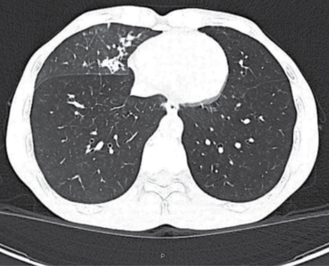
Male, 52 years old, resident of Chimalhuacán, State of Mexico, bricklayer, history of visiting caves in Veracruz; no chronic degenerative diseases, cancer, use of drugs, denies smoking and ethylism. He started two years before, with cough with yellowish expectoration, occasional hemoptoics without hemodynamic or respiratory repercussions; weight loss of 22 kilos in six months, asthenia, adynamia, dysphagia to solids, as well as constant holocranial headache without identifying exacerbating or exacerbating factors. With loss of alertness 20 days before his admission, he went to the doctor who requested a tomographic study in which a lymph node conglomerate was observed at paratracheal 4R and subcarinal 7, at the level of the middle mediastinum a heterogeneous image of 4.7 × 4.8 cm with 75 HU and contrast enhancement, extrinsic compression of the esophagus and alveolar filling pattern in the middle lobe associated with solid and semi-solid spiculated nodules was observed (Figure 1).
At the outpatient clinic, bronchoscopy was performed with findings of irregular mucosa with nodular lesions in the right main bronchus extending to the bronchus intermedius, cryobiopsy of bronchus intermedius and bronchioalveolar lavage of the right lower lobe, with positive Grocott and Schiff's periodic acid, compatible with Cryptococcus sp., without presence of malignant cells, negative GeneXpert. Panendoscopy was performed with extrinsic compression of the middle esophagus, retentive stomach and erosive gastropathy, being negative for microorganisms and neoplastic cells in histopathology. He was hospitalized to continue the approach and start antifungal treatment. Physical examination showed no neurological alterations, chest with diminished respiratory sounds in the right subscapular region; hemogram, blood count, blood sugar, glycated hemoglobin in normal ranges; no transaminasemia or hyperbilirubinemia, negative inflammatory markers, serology for human immunodeficiency virus (HIV), hepatitis B virus (HBV) and hepatitis C virus (HCV) were not reactive.
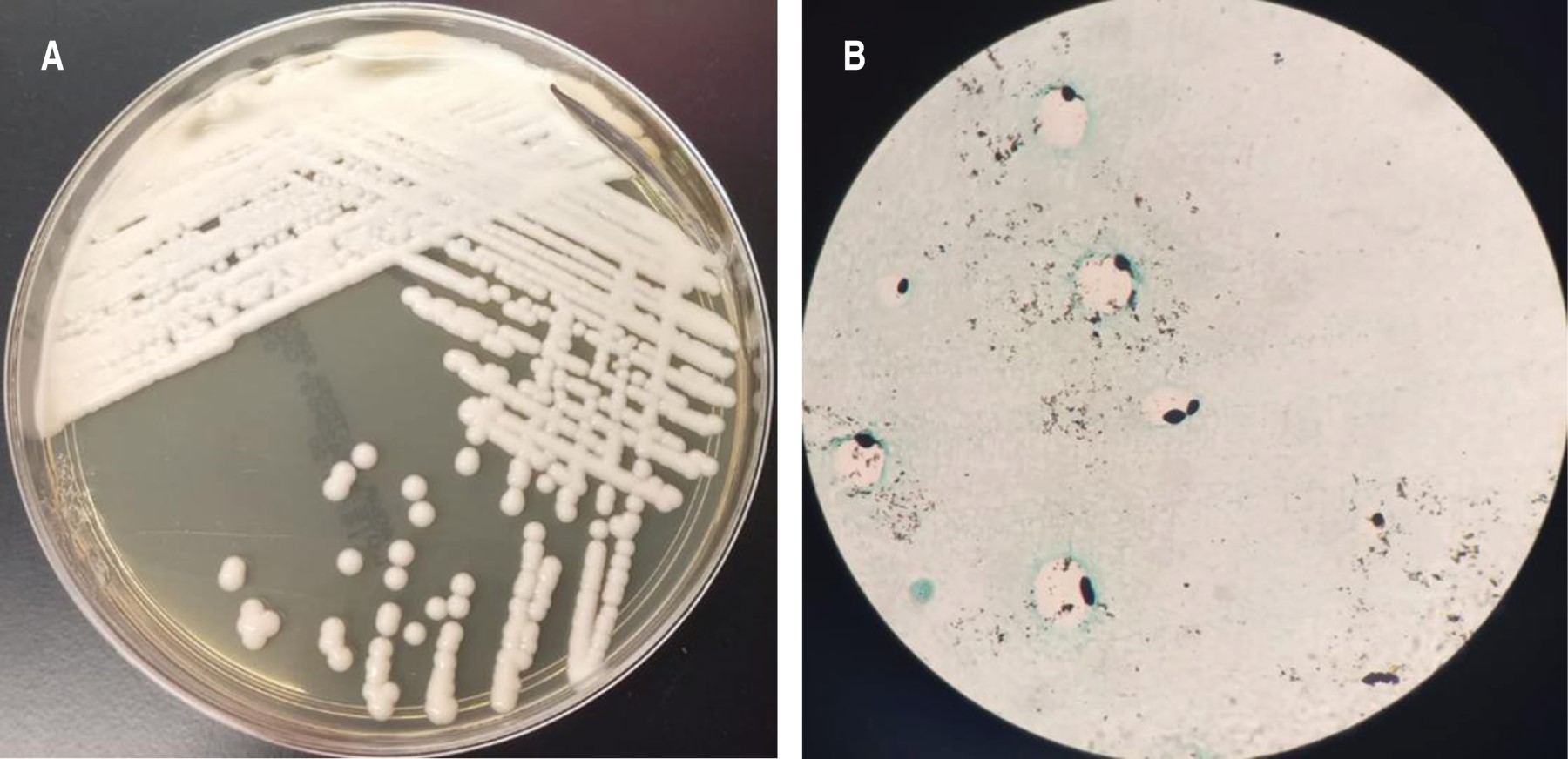
During hospitalization, a tomographic study of the brain was performed, but no alterations were found; a lumbar puncture was performed with positive results in the meningitis panel for Cryptococcus, positive Chinese ink and Grocott, positive culture result for Cryptococcus gattii (Figure 2). A week after admission, a lung biopsy was performed by videothoracoscopic surgery (VATS) of the lower lobe lesion in segment 6 and upper lobe in segment 3, with negative trans-operative result for malignancy, pericardial infiltration of the mediastinal lesion was found, and a pericardial window was performed. Histopathology in lung parenchyma sections showed pneumonia in various stages of organization without evidence of microorganisms, pericardium and mediastinum sections with spherical structures of reinforced wall, with pseudohypha and clear peripheral halo positive for Schiff and Grocott periodic acid stains, concluding cryptococcosis, Ziehl-Neelsen stain negative. After 20 days he presented again with pericardial effusion with 28 mm separation and pleural effusion, washing and drainage of the thoracic cavity plus pericardial window was performed, stains, pericardial biopsy culture and pericardial fluid were negative. Treatment with fluconazole and liposomal amphotericin B was maintained. Outpatient follow-up is planned to rule out primary immunodeficiency or other added factors.
Discussion
Cryptococcus is a genus of encapsulated yeasts belonging to the phylum Basidiomycota and is widely distributed worldwide. Several forms of presentation have been described, the most frequent being pulmonary in its localized form and meningeal in its disseminated form. The main route of entry is inhalation, with easy dissemination by the hematogenous route with great tropism for the central nervous system.3
Pulmonary cryptococcosis in an immunocompetent patient is rare. However, a retrospective review in China reported that 60% of pulmonary cryptococcosis cases were diagnosed in immunocompetent patients.4 Its spread is even rarer, which can lead to fatal complications, with mortality being even higher than those with underlying HIV infection, making early diagnosis critical.5 A previous study reported that 67% of pulmonary cryptococcosis in immunocompetent patients spread to the central nervous system causing cryptococcal meningitis.6
Both the innate and adaptive immune systems participate in the host response against Cryptococcus infection. Alveolar macrophages phagocytize Cryptococcus, and a granulomatous immune response may follow with the formation of a subpleural nodule or primary lymph node, with pulmonary nodules being a common manifestation on radiological examination. The fate of Cryptococcus within macrophages is determined by the immune status of the host, if compromised it can lead to reduced pulmonary clearance and possible dissemination.7 In those who are immunocompetent, it has been reported that those with pulmonary cryptococcosis have a defect in their immune system, such as the polymorphism in Dectin-2, studied by Hu in China.8
Recognition of the disease in immunocompetent patients can be difficult, as the presentation is often more indolent and subtle, like the presenting patient. This can result in delayed diagnosis and initiation of treatment, which can lead to complications and the development of more severe disease. Some studies have reported higher mortality in non-HIV-infected patients than in HIV-infected patients.9 Initiation of antifungal therapy is important for prevention of complications, which can be particularly challenging in this population.
Conclusions
This report documents a rare case of disseminated cryptococcosis due to Cryptococcus gattii in an immunocompetent patient. We emphasize the importance of knowing the different forms of presentation, as well as early diagnosis and timely treatment to reduce the risk of complications and sequelae.
AFILIACIONES
1Instituto Nacional de Enfermedades Respiratorias Ismael Cosío Villegas, Mexico City, Mexico.Conflict of interests: the authors declare that they have no conflict of interests.
REFERENCES
Suwatanapongched T, Sangsatra W, Boonsarngsuk V, Watcharananan SP, Incharoen P. Clinical and radiologic manifestations of pulmonary cryptococcosis in immunocompetent patients and their outcomes after treatment. Diagn Interv Radiol. 2013;19(6):438-446. Available in: https://doi.org/10.5152/dir.2013.13049


