Symmetrical bilateral effusion and spondylodiscitis: a case report
Tovar-Becerra, Andrea Itzel1; García-Hernández, Jorge A1; Sosa-Hernández, Oscar1
Tovar-Becerra, Andrea Itzel1; García-Hernández, Jorge A1; Sosa-Hernández, Oscar1
ABSTRACT
KEYWORDS
symmetrical bilateral effusion, spondylodiscitis, extrapulmonary tuberculosis.Introduction
Empyema is defined as the presence of purulent fluid in the pleural cavity, which is mostly associated with infections of the pulmonary parenchyma, where pneumonia is the most frequent; other etiological factors are trauma, esophageal rupture, after thoracic surgery or infections of the cervical and thoracic spine.1
The presentation of bilateral empyema is rare and there are no guidelines that establish the systematic approach or management. The evaluation of these patients is challenging because the differential diagnosis is broad and includes both benign and malignant, and even life-threatening conditions.2 Empyema is associated with an increased risk of mortality and therefore requires timely, multidisciplinary intervention.
The following clinical case of symmetrical bilateral empyema associated with spondylodiscitis is presented, in which symptomatology, radiological findings and epidemiological aspects (demography, socioeconomic status, malnutrition) are correlated to reach the final diagnosis in a timely manner in order to reduce morbimortality.
Case presentation
Male, 60 years old, work history in the chemical materials and mining industry, systemic arterial hypertension for eight years of diagnosis, alcoholism for 30 years, smoking for 13 years, smoking rate 6.5, exposure to biomass for 11 years for three hours a day.
Two months of evolution with stabbing pain VAS 9/10 in the right costal region, self-medication with non-steroidal anti-inflammatory drugs (NSAID) without improvement; adding fever, asthenia, adynamia, hyporexia, oral thrush, involuntary weight loss (10 kg), nocturnal diaphoresis, dysphagia and dry cough. One week prior to her admission, she presented weakness in the pelvic limbs with difficulty in standing and inability to ambulate without alteration of sensibility.
On physical examination, the patient was found cachectic, malnourished, body mass index (BMI) 18, chest stethoacoustic, heart sounds without alterations, pulmonary area integrated in posterior region, bilateral basal pleural effusion syndrome in 50%, pelvic limbs hypotrophic, preserved sensitivity, but decreased mobility and strength, Daniels scale 2/5 left and 3/5 in right, respectively, with inability to ambulation, myotatic ++/+++, without data of pyramidal release, the rest of the physical examination without alterations.
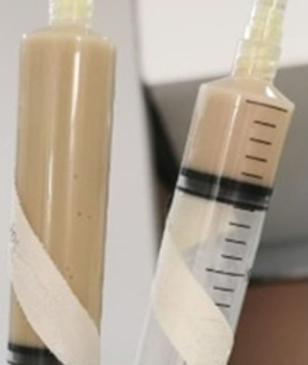
Blood cytometry documented leukocytosis 20,700 thousands/UL at the expense of polymorphonuclear (PMN) 16,170 cells, lactate dehydrogenase 122 U/L, albumin 2.4 g/dL, C-reactive protein 28.9 mg/dL. Thoracentesis was performed which revealed fetid purulent fluid (Figure 1) documenting cellularity 324,000 mm3, polymorphonuclear 81%, mononuclear 19%, erythrocytes 23,500 million/UL, crenated 45%, non-crenated 55%, pH 5, glucose 45 mg/dL, lactate dehydrogenase 20,376 U/L, total protein 2. 90 g/dL, albumin 0.05 g/dL, cholesterol 387.50 mg/dL, total bilirubin 0.180 mg/dL.
Magnetic resonance imaging (MRI) was performed, which documented, in pleural space, oval images of hypointense interior in T1, hyperintense T2 and SPAIR with wall thickening up to 11 mm, presented communication with destroyed vertebral body in T12, which presented changes due to spondylodiscitis that conditioned spinal cord contact and increase in its amplitude without change in intensity (Figure 2).
Histopathological report of pleural fluid: yellowish-greenish and fetid fluid is described; in the microscopic description, on a protein background and with necrosis, inflammatory cells, lymphocytes in moderate quantity and abundant neutrophils are observed. Negative for neoplasia, intense acute and chronic inflammation. Pleural fluid culture negative. Gram stain showed no bacterial forms and negative Ziehl-Neelsen stain. As part of the empyema management, bilateral endopleural probe placement was performed without complications, being the evolution of the patient unfavorable and torpid, so the case was discussed with neurosurgery for vertebral lesion approach. However, due to the patient's condition, he did not receive a surgical procedure and was evaluated in conjunction with the Epidemiology Service, which, given the clinical manifestations and the negative biochemical reports for bacterial and neoplastic development, in addition to the MRI findings, considered the possibility of tuberculous etiology and decided to initiate antituberculosis treatment, presenting improvement in his condition and a decrease in acute phase reactants.
The neurosurgical approach to take a biopsy of the vertebral lesion was left pending until the patient's clinical condition allowed it.
Discussion
The incidence of empyema is variable, with 32,000 cases reported per year in the United States (US); however, there are reports of increasing frequency.3 It has been reported that up to 40% of patients with community-acquired pneumonia will develop pleural effusion; and of these, up to 7% will develop complicated parapneumonic effusion or empyema. The bacterial cause is the most frequent; Falguera et al. described that, of the 261 patients with empyema, 64% were gram-positive cocci, 6% gram-negative, 10% anaerobic and 4% atypical microorganisms.4
Independent risk factors for the development of empyema include diabetes, immunosuppression, gastroesophageal reflux disease, aspiration and poor oral hygiene, alcohol and intravenous drug abuse.4
Our case reports a rare association of bilateral empyema and spondylodiscitis. Pleural fluid analysis confirmed exudative features; although pleural fluid cultures did not report development, cytology did not document neoplastic cells and even the search for BAAR was negative. Due to unfavorable clinical conditions the patient was not a candidate for bone biopsy.
In 2021, San Luis potosí, Mexico, ranked seventh nationally in prevalence of extrapulmonary tuberculosis (EPTB), reporting 82 cases.5 The diagnosis of EPTB is complicated and should be suspected on an epidemiological basis in countries with high prevalence and/or lack of response to conventional treatment.
The simultaneous presentation of vertebral tuberculosis (VTB) with pleural involvement is infrequent, occurring in about 2.5% of patients.6 Some authors have reported that 10% of extrapulmonary forms correspond to osteoarticular tuberculosis, of which up to half have VTB.6 In the USA and Europe it represents 10-15% and 2-4.7%, respectively, of all cases of tuberculosis.7 The lower thoracic and lumbar vertebrae are the most common sites of involvement.8 The mechanism of infection is considered to be a primary focus at the vertebral level with direct dissemination by contiguity to the pleura. This suggests an atypical natural history of the disease.6
The diagnosis of VTB is complex due to the low specificity of clinical and paraclinical data, so it must be based on clinical and epidemiological correlation and radiological findings, with magnetic resonance imaging being the study of choice.9 The delay in the diagnosis of vertebral involvement leads to neurological complications between 30-80% due to spinal cord involvement, which increases morbidity and mortality.
Considering the aforementioned, the possibility of tuberculous etiology was considered, based on the endemic behavior of this mycobacterium in our country and the adequate response and improvement of the symptomatology with decrease of the systemic inflammatory response to the antituberculosis treatment.
Our case highlights an unusual presentation of bilateral empyema associated with spondylodiscitis, where the imaging findings in a patient with risk factors living in an endemic area for tuberculosis were determinant to consider the diagnosis and initiate a specific treatment.
Conclusions
Tuberculosis should be considered as a rule-out diagnosis in cases of empyema in patients with or without risk factors and living in endemic areas, especially if associated with vertebral lesions. Management and diagnosis should be multidisciplinary, and this is the justification for initiating antituberculosis treatment. The support of imaging studies allows timely diagnosis and treatment to avoid complications and improve the quality of life of patients.
Acknowledgments
To Dr. Cuellar for his valuable participation in the interpretation of cabinet studies. To Dr. Helena Rodríguez for her contributions.
AFILIACIONES
1Hospital General de Zona No. 50, Instituto Mexicano del Seguro Social. San Luis Potosí, Mexico.Conflict of interests: the authors declare that they have no conflict of interests.
REFERENCES


