
Introduction
COVID-19 can progress to acute respiratory distress syndrome (ARDS), characterized by alveolar collapse, exudative consolidation, and pulmonary inflammation that may progress to biotrauma and multiple organ failure. In this context, mechanical ventilation is an essential supportive therapy that maintains gas exchange while allowing the lung to recover, through careful adjustment of volume and pressure to optimize ventilation and promote alveolar recruitment.
Protective ventilation, based on tidal volumes of 6-8 ml/kg of predicted body weight, aims to avoid both overdistension (volutrauma) and cyclic alveolar collapse (atelectrauma), which is particularly relevant in ARDS, where lung injury is already established.1 This condition compels the mechanical ventilator operator to exercise extreme caution in selecting administered volumes and pressures, with the goal of neither perpetuating nor worsening the underlying lung injury. The settings used in this context are known as lung-protective parameters.
The indication for invasive mechanical ventilation (IMV) in patients with COVID-19 is established when severe respiratory failure is refractory to other therapeutic strategies, such as conventional oxygen therapy, non-invasive mechanical ventilation, or high-flow nasal cannula. In particular, IMV should be considered when one or more of the following criteria are met: a) arterial oxygen pressure (PaO2)/inspired oxygen fraction (FiO2) ratio < 200; b) deterioration of neurological status; c) increased work of breathing (WOB); d) tachypnea greater than 30 breaths per minute; and e) respiratory acidosis, defined as pH < 7.35 with PaCO2 > 45 mmHg2 at altitudes such as that of Mexico City at 2,240 m. To adequately understand ventilatory parameters and monitoring strategies, it is essential to be familiar with the clinical phenotypes described throughout the pandemic.
COVID-19 phenotypes
Patients with COVID-19 may present with different patterns of refractory hypoxemia, ranging from cases with preserved pulmonary compliance to more severe forms resembling classic ARDS. Some patients show a good response to prone positioning, while others without improvement tend to have low alveolar recruitment capacity.3 Gattinoni et al.,3 in their editorial COVID-19 pneumonia: different respiratory treatments for different phenotypes?, hypothesize that the clinical presentation and severity of COVID-19 pneumonia depend on the interaction of multiple factors: 1) the severity of infection, the host response, physiological reserve, and comorbidities; 2) the ventilatory response to hypoxemia; and 3) the interval between symptom onset and initiation of hospital management.
Two main phenotypes have been proposed:
-
− Phenotype L is characterized by relatively preserved compliance, predominantly perfusion-related ventilation/perfusion mismatch, a ground-glass pattern with low lung weight, and limited recruitment response.
− Phenotype H, in contrast, presents with high elastance, increased edema and lung weight, greater intrapulmonary shunt, and high alveolar recruitment capacity. It resembles classic ARDS.3
These phenotypes can evolve from L to H due to increased ventilatory work and intrathoracic pressures, as well as progression of inflammatory injury and edema. This pathophysiological dynamic explains the clinical variability observed in severe COVID-19, including heterogeneous responses to ventilation and oxygenation.3 Figure 1 illustrates the differences between the two phenotypes.
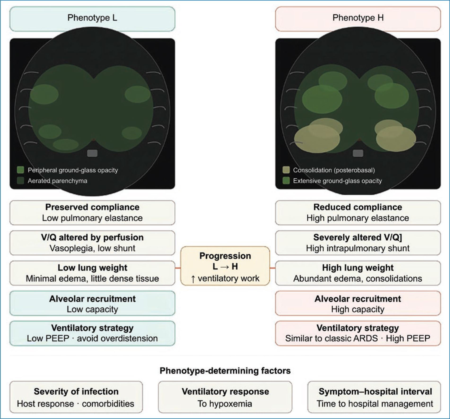
Figure 1. Phenotypes of severe COVID-19 pneumonia. Schematic axial cross-sections illustrate the characteristic computed tomography findings of each phenotype. Phenotype L (left panel) shows peripheral and sparse ground-glass opacities, with predominantly aerated pulmonary parenchyma and low lung weight. Phenotype H (right panel) presents extensive posterobasal consolidations and diffuse ground-glass opacities, reflecting abundant pulmonary edema and increased lung weight. Physiologically, phenotype L is characterized by preserved compliance, predominantly vascular ventilation/perfusion mismatch, and low recruitment capacity; phenotype H, in contrast, resembles classic ARDS, with high elastance, elevated intrapulmonary shunt, and high alveolar recruitment capacity. Both phenotypes may coexist or progress from L to H due to increased ventilatory work and progression of inflammatory injury. The images are schematic representations; they do not correspond to real patient computed tomography scans. Adapted from Gattinoni et al.3. PEEP: positive end-expiratory pressure; ARDS: acute respiratory distress syndrome; V/Q: ventilation/perfusion ratio.
The H and L phenotypes described by Gattinoni constitute a useful conceptual tool for understanding the pathophysiological heterogeneity of COVID-19; however, hyperinflammatory and hypoinflammatory sub-phenotypes of ARDS have since been described, making individual assessment of pulmonary mechanics, recruitability, and clinical response is of greater relevance for guiding therapeutic decisions.
Initial ventilator settings in mechanical ventilation
Initial mechanical ventilation programming should be individualized according to the patient’s clinical characteristics, with emphasis on lung protection, oxygenation, and ventilator synchrony. No single ventilatory mode is superior, so the choice depends on clinical experience. However, it is frequently recommended to begin with volume-controlled ventilation to ensure a protective tidal volume (VT) (4-8 ml/kg of predicted body weight, ideally 6-8 ml/kg), with a respiratory rate of 12-18 breaths/min and adjustments that may require increments according to the patient’s condition.4
Other initial parameters include an inspiratory flow of 40-60 l/min, an inspiratory-to-expiratory ratio (I:E) of 1:2 to 1:3, an initial positive end-expiratory pressure (PEEP) ≥ 5 cmH2O, and a sensitivity of 1.5 to 2 l/min (or, in pressure-triggered mode, between −1 and −3 cmH2O). The central objective is to maintain a plateau pressure (Pplat) < 30 cmH2O and to avoid ventilator-induced lung injury (VILI) by simultaneously adjusting rate and flow to prevent auto-PEEP. Minute volume must be adapted to metabolic demands and dead space. In some cases, permissive hypercapnia may be accepted as a protective strategy.2
Pressure-controlled mode is a valid alternative, but it requires knowledge of its programming differences depending on the ventilator. In this mode, VT is variable and depends on the patient’s pulmonary mechanics and the applied pressure gradient, such that thoracopulmonary impedance directly influences effective ventilation; with greater impedance, peak inspiratory pressure is reached more rapidly, resulting in a lower delivered VT. From an operational standpoint, the pressure difference (ΔP) between the baseline (PEEP plus auto-PEEP) and peak inspiratory pressure (PIP) is the primary determinant of the administered VT. This relationship is influenced by both the mechanical properties of the lung and the patient’s respiratory effort.4 Other relevant considerations include the use of a preferentially decelerating flow waveform, programming a minimum respiratory rate, appropriate adjustment of the I:E ratio, and trigger sensitivity. Inspiratory pressure should be titrated progressively to achieve the target tidal volume within a safe range, in accordance with the principles of lung protection.4
Concept of recruitment
Pulmonary recruitment, described by Lachmann in 1992, consists of the reopening of collapsed alveolar units to restore them to ventilation. This process is achieved through the transient application of elevated inspiratory pressures (positive inspiratory pressure) and their maintenance with adequate PEEP levels, in order to prevent end-expiratory collapse and promote more homogeneous and protective ventilation.5 Alveolar recruitment maneuvers aim to increase transpulmonary pressure in a controlled manner to reopen collapsed or partially ventilated alveoli. In the context of ARDS, they represent a relevant strategy in the management of refractory hypoxemia. However, the available evidence, particularly in COVID-19, is limited and of low quality, so their use is not recommended systematically. When employed, they may include the application of elevated pressures over brief periods, always under close clinical monitoring.6,7,8,9
Pulmonary monitoring
The interpretation of pulmonary mechanics variables is fundamental to understanding the functional state of the lung and guiding mechanical ventilation adjustments. It is necessary to measure, analyze, and interpret the forces applied during insufflation – particularly transpulmonary pressure (stress) and the resulting deformation of pulmonary tissue (strain) – without losing sight of the central objective of maintaining open and functional alveolar units for adequate gas exchange. From the perspective of the ventilation/perfusion (V/Q) relationship, ventilation can be understood as the response of the respiratory system to the mechanical impulse of insufflation and its subsequent return to the resting position. This process is divided into three components: 1) the dynamic phase, in which the resistive forces of the respiratory system (lung, thoracic cage, and abdomen) are overcome; 2) the static phase, corresponding to the distribution of gas within pulmonary compartments; and 3) the elastic recoil phase of the lung. Identifying the variables that represent each of these phases is essential for adequate monitoring and for titrating a protective ventilatory strategy.6,7,8,9
Monitoring under static conditions
In volume-controlled mechanical ventilation, assessment under zero-flow conditions allows the elastic properties of the respiratory system to be analyzed with greater precision. During the inspiratory pause – after closure of the inspiratory valve and before opening of the expiratory valve – flow ceases, and the administered volume redistributes homogeneously. At this point, the plateau pressure (Pplat) is measured, which reflects the elastic recoil pressure of the respiratory system and, in equilibrium, approximates alveolar pressure.8
Additionally, intrinsic PEEP (PEEPi) is measured through an end-expiratory pause, also under zero-flow conditions, allowing the identification of air trapping and dynamic hyperinflation. PEEPi may occur in different scenarios: 1) without pulmonary overdistension, associated with active expiration and increased end-expiratory alveolar pressure; 2) due to increased ventilatory demands (elevated respiratory rate or tidal volume) with insufficient expiratory time, leading to incomplete alveolar emptying; and 3) in the presence of airflow limitation, where dynamic airway collapse during expiration promotes air trapping and increased alveolar pressure without improvement in expiratory flow.10 Correct interpretation of these variables is essential for optimizing ventilation and minimizing the risk of ventilator-induced lung injury.
Compliance (C) describes the relationship between the change in lung volume and the pressure required to generate it (C = V/P, ml/cmH2O). In patients under IMV, static compliance is estimated from the tidal volume (VT) divided by the difference between Pplat and PEEP (C = VT/(Pplat−PEEP)). As a reference, normal values are approximately 100 ml/cmH2O under general conditions, approximately 75 ml/cmH2O in the supine position, and approximately 50 ml/cmH2O in patients with ARDS, reflecting the loss of pulmonary elasticity in this context.11
Driving pressure is defined as the difference between Pplat and PEEP and is ideally evaluated in the absence of spontaneous respiratory effort. This parameter reflects the relationship between tidal volume and respiratory system compliance, serving as an indirect indicator of functional lung size. Clinical evidence has demonstrated that elevated driving pressure is associated with higher mortality in patients with ARDS; therefore, it is recommended to maintain it below 15 cmH2O. Furthermore, monitoring this parameter is useful for adjusting PEEP within a protective ventilation strategy aimed at optimizing oxygenation and minimizing lung injury.12
Monitoring under dynamic conditions
Dynamic monitoring allows evaluation of the interaction between flow, pressure, and volume during the respiratory cycle, integrating both the elastic and resistive properties of the respiratory system.
Airway resistance is defined as the relationship between the pressure gradient (from the proximal airway to the alveoli) and airflow (R = P/F, cmH2O/L/s). This parameter varies as a function of lung volume: the greater the volume, the lower the airway resistance, and vice versa.13 Its assessment is useful for identifying airflow obstruction or changes in the mechanical conditions of the respiratory system.
Dynamic compliance is calculated as the ratio between VT and the difference between peak pressure and PEEP (Cdyn = VT/(Ppeak−PEEP)). Unlike static compliance, this parameter integrates the combined effect of airway resistance, the thoracic cage, and the pulmonary parenchyma. Its values are typically 10-20% lower than those of static compliance and may be influenced by factors such as the patient’s age and weight.13
Monitoring of pulmonary parenchymal deformation
Pulmonary stress is defined as the distending pressure applied to the fibro-skeleton of the lung during ventilation. Based on this concept, Grasso et al.14 developed the stress index, a useful tool for optimizing PEEP titration and reducing the risk of VILI. This index is evaluated through the morphology of the pressure-time curve (Fig. 2). A downward-concave curve (index < 1) indicates increased compliance and suggests potential alveolar recruitment, such that increasing PEEP to achieve a more linear tracing might be considered. In contrast, an upward-concave curve (index > 1) suggests alveolar overdistension, indicating the need to reduce PEEP. A linear profile is associated with more homogeneous ventilation and, therefore, with a lower risk of lung injury.
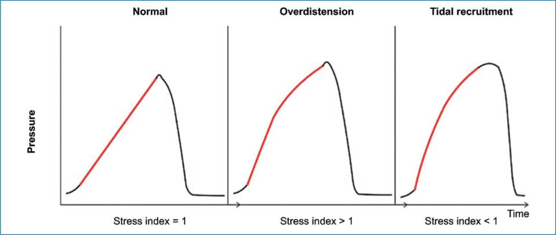
Figure 2. Monitoring of the stress index on the pressure curve (y-axis) and time (x-axis) during invasive mechanical ventilation. The red segment marks the inspiratory portion in each case: Normal (= 1): pressure rises linearly during inspiration; Overdistension (> 1): the curve accelerates (upward concavity), indicating alveolar overdistension; Tidal recruitment (< 1): the curve decelerates (downward concavity), indicating cyclic recruitment with each breath.
Strain represents the deformation of the pulmonary fibroskeleton in response to mechanical ventilation and constitutes a key parameter in the genesis of VILI. Its assessment allows estimation of the degree of mechanical stress applied to the lung and guides protective ventilation strategies. Among the available tools, electrical impedance tomography stands out as a non-invasive, radiation-free, and low-cost method that allows real-time analysis of the regional distribution of ventilation, contributing to optimization of ventilatory parameters and reduction of VILI risk.14
Ventilator titration and lung protection
Mechanical ventilation titration is directed toward three fundamental objectives: avoiding alveolar overdistension, optimizing alveolar recruitment (preventing cyclic collapse and reopening), and adjusting ventilation to the characteristics of the “injured lung,” in accordance with oxygenation and ventilation targets.15
Establishing a VT based on predicted body weight constitutes the cornerstone of protective ventilation. Evidence has demonstrated that the use of low VT (≈6 ml/kg) is associated with lower mortality compared with higher volumes (≈12 ml/kg), primarily due to its impact on reducing plateau pressure.16,17 Nevertheless, in patients with COVID-19 phenotype L, characterized by greater compliance, the use of slightly higher volumes (up to 8 ml/kg of predicted body weight) may be considered, provided that plateau pressure remains below 30 cmH2O.18
Maintaining a plateau pressure < 30 cmH2O is a key objective, as this parameter correlates with alveolar distension and, under normal conditions, acts as a surrogate for transpulmonary pressure.14 However, its interpretation must be adjusted in the presence of alterations in thoracic or abdominal elastance, where it may lose precision and require more direct measurements, such as esophageal pressure. Elevated plateau pressure values are associated with a greater risk of alveolar edema and lung injury.2
Driving pressure (ΔP = plateau pressure−PEEP) reflects the available functional lung volume and the degree of recruitable lung in each respiratory cycle. This parameter integrates both the ventilator configuration and the patient’s mechanical properties, and it is recommended to maintain it below 15 cmH2O as part of a protective ventilation strategy.13,16
Importance of PEEP titration in patients with COVID-19
Adequate PEEP titration is a key component of the protective ventilation strategy, particularly in patients with ARDS secondary to COVID-19. A meta-analysis that included the ALVEOLI, LOVS, and EXPRESS studies evaluated the impact of high versus low PEEP strategies.19 In the overall analyzed population, no significant differences in mortality were observed between the two groups (relative risk [RR]: 0.94; 95% confidence interval [95% CI]: 0.86-1.04; p = 0.25). However, in the subgroup of patients with ARDS, the use of higher PEEP levels was associated with a modest but significant reduction in mortality (adjusted RR: 0.90; 95% CI: 0.81-1.0; p = 0.049). These findings suggest that the benefit of elevated PEEP depends on the severity of the syndrome. In particular, patients with moderate to severe ARDS may benefit from higher PEEP levels, likely due to greater alveolar recruitment potential. In contrast, in patients with mild ARDS, the use of elevated PEEP not only lacks clear benefit but could also be associated with adverse effects, including alveolar overdistension and hemodynamic compromise. Therefore, PEEP titration must be individualized, considering the clinical response, pulmonary mechanics, and the recruitment potential of each patient.20
Prone positioning
In ARDS, areas of alveolar collapse and consolidation predominate in the dependent regions of the lung. In the supine position, these correspond primarily to the dorsobasal regions, promoting ventilation-perfusion imbalance. Ventilation in the prone position redistributes these areas, shifting the collapse toward anterior regions and promoting more homogeneous ventilation of the pulmonary parenchyma.21
This change improves the ventilation/perfusion ratio, reduces intrapulmonary shunt, and decreases both alveolar overdistension and dead space, thereby helping to limit VILI.21 The benefits are most evident in patients with moderate to severe ARDS, in whom a significant reduction in mortality has been demonstrated, as evidenced by the PROSEVA study.22 Table 1 presents some characteristics of the studies that have employed this therapeutic strategy.
Table 1. Methodological aspects of studies that have implemented prone positioning ventilation in patients with ARDS
| Characteristic | Classic ARDS | COVID-19 ARDS |
|---|---|---|
| Study | Guérin et al.22 PROSEVA trial | Observational studies and meta-analyses (2020-2024) |
| Study design | Multicenter randomized clinical trial | Predominantly observational (cohort, retrospective, meta-analyses) |
| Level of evidence | High | Moderate-low |
| Population | Severe ARDS (PaO2/FiO2 < 150) | Moderate-severe ARDS, heterogeneous population |
| Pathophysiology | “Typical” ARDS | Initially variable phenotypes; currently managed as typical ARDS |
| Onset of prone | Early (< 36 hours) | Variable; often delayed at the start of the pandemic |
| Duration of prone | ≥ 16 hours per session | ≥ 16 hours; frequently prolonged (> 24 hours) |
| Number of sessions | Repeated cycles | Variable according to clinical response |
| Ventilatory strategy | Strict protective ventilation | Variable adherence to protective ventilation |
| Effect on oxygenation | Significant and consistent improvement in PaO2/FiO2 | Consistent improvement, with variable magnitude |
| Effect on mortality | Significant reduction (28 days: 16% versus 32.8%) | Inconclusive; possible benefit suggested |
| Complications | Pressure ulcers, airway events (without significant increase) | Similar profile; manageable in experienced centers |
| Reproducibility | High (protocolized intervention) | Variable (resource- and experience-dependent) |
| Clinical recommendation | Strong recommendation | Strong recommendation (based on extrapolation and physiological benefit) |
ARDS: acute respiratory distress syndrome.
In the context of COVID-19, although distinct phenotypes were initially proposed, current evidence supports that ventilatory management (including prone positioning) should follow the principles of classic ARDS. In these patients, especially those with severe hypoxemia (PaO2/FiO2 < 150, FiO2 > 60%, and PEEP ≥ 5-10 cmH2O), prone positioning consistently improves oxygenation and is associated with better outcomes when applied early and for prolonged periods. To maximize its benefits, it is recommended to maintain the prone position for > 12 hours per day,23 ideally during the exudative phase of ARDS and under adequate sedation. More recent evidence suggests that extending sessions beyond 24 hours may be associated with greater benefits in terms of oxygenation and clinical outcomes;24 however, this approach must be balanced against the risk of complications, most notably pressure ulcers and other injuries associated with prolonged immobility.25,26 Discontinuation of this strategy should be considered when sustained improvement in oxygenation is achieved (PaO2/FiO2 > 150, FiO2 < 60%, and PEEP < 10 cmH2O), with stability verified after returning to the supine position. In the event of deterioration, resumption of prone positioning should be reassessed.27
Contraindications include significant hemodynamic instability or conditions that make its implementation difficult, such as recent abdominal surgery or cranioencephalic trauma. Its application likewise requires a trained multidisciplinary team, given the level of complexity and resources required.28 Currently, the evidence supports its use as a standard intervention in the management of ARDS, regardless of the cause, including COVID-19.23
Prone positioning constitutes a fundamental strategy in the management of intubated patients with COVID-19 and severe ARDS. Its timely and appropriate implementation, particularly in intensive care units, has proven to be a key intervention for improving oxygenation and clinical outcomes, as shown in figure 3.
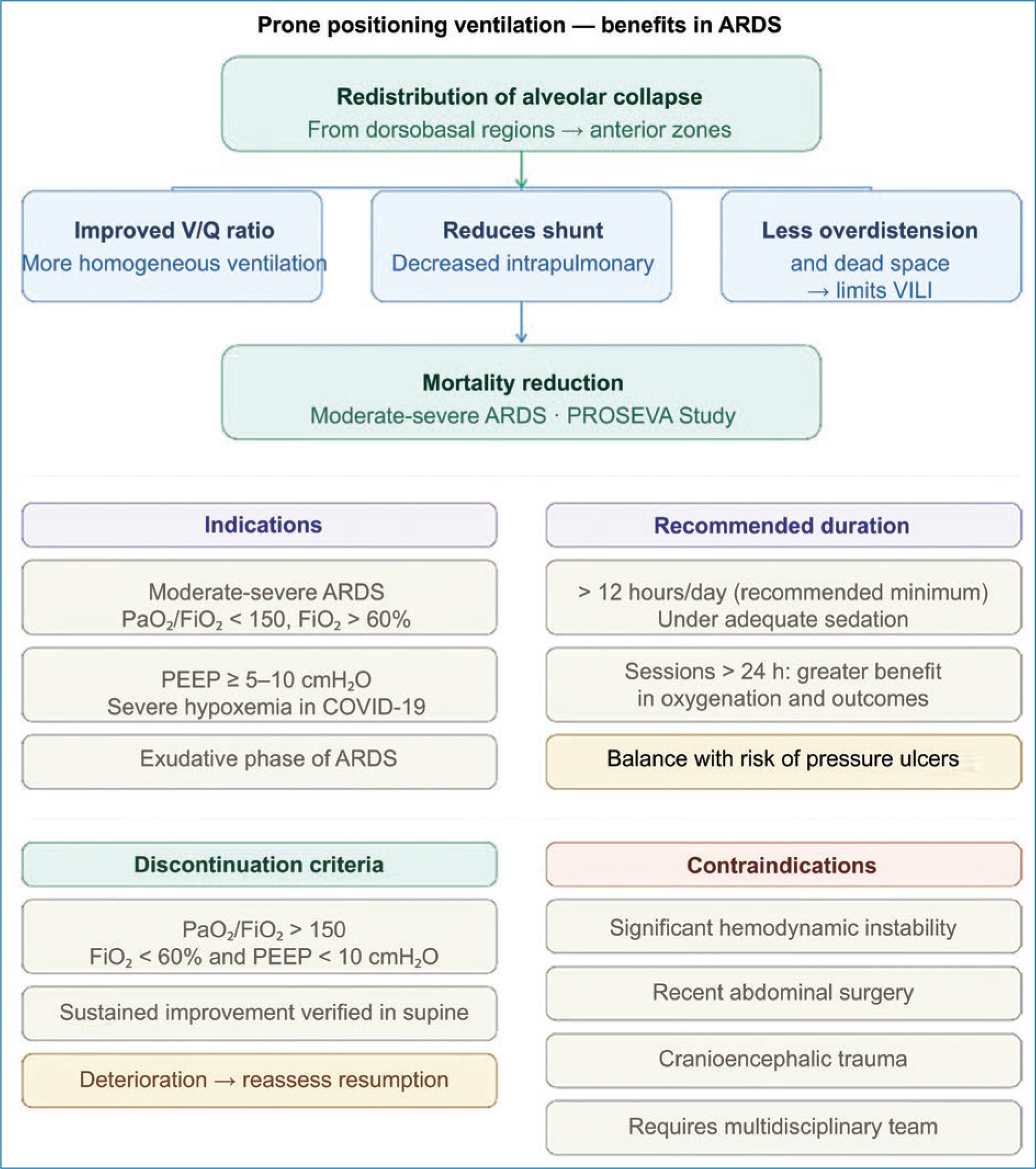
Figure 3. Physiological benefits and clinical considerations of prone positioning ventilation in acute respiratory distress syndrome (ARDS). The postural change redistributes alveolar collapse from the dorsobasal regions toward anterior zones, promoting more homogeneous ventilation of the pulmonary parenchyma. This results in improved ventilation/perfusion (V/Q) ratio, reduced intrapulmonary shunt, and decreased ventilator-associated lung injury (VILI). PEEP: positive end-expiratory pressure; ARDS: acute respiratory distress syndrome; V/Q: ventilation/perfusion ratio; VILI: ventilator-induced lung injury.
Conclusion
Taken together, current evidence confirms that ventilatory management of ARDS – including that associated with COVID-19 – should be grounded in a comprehensive protective ventilation strategy, based on limiting ventilator-induced injury, optimizing alveolar recruitment, and individualizing parameters according to each patient’s pulmonary mechanics. Continuous monitoring of static and dynamic variables, alongside advanced regional assessment tools, allows precise ventilation adjustments to minimize VILI. Likewise, interventions such as appropriate PEEP titration and prone positioning continue to play a central role in improving oxygenation and reducing mortality in moderate to severe cases. In this context, the pathophysiological understanding of ARDS and the rigorous application of lung protection principles remain fundamental pillars for optimizing clinical outcomes in critically ill patients.
Funding
The present work received no funding.
Conflicts of interest
The authors declare no conflicts of interest.
Ethical considerations
Protection of persons and animals. The authors declare that no experiments were performed on human beings or animals for this research.
Confidentiality, informed consent, and ethical approval. The study does not involve personal patient data and does not require ethical approval. The SAGER guidelines do not apply.
Statement on the use of artificial intelligence. The authors declare that they did not use any form of generative artificial intelligence in the writing of this article.
